


Digestive System of the Pig: Anatomy and Function
An overview of the pig's digestive system - mouth, stomach, small and large intestines by Joel DeRouchey and colleagues at Kansas State University's Applied Swine Nutrition Team, presented at the Swine Profitability Conference 2009. 10 June 2009
 6 minute read
The digestive system of a pig is well suited for complete concentrate based rations that are typically fed. The entire digestive tract is relatively simple in terms of the organs involved, which are connected in a continuous musculo-membanous tube from mouth to anus. Yet this multi-faceted system involves many complex interactive functions.
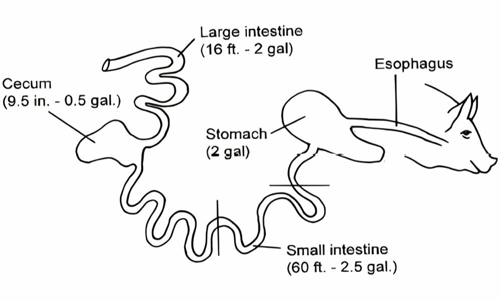
The goal of this paper is to describe the organs involved in digestive and biological functions (Figure 1).
Mouth
The mouth serves a valuable role not only for the consumption of food but it also provides for the initial partial size reduction though grinding. While teeth serve the main role in grinding to reduce food size and increase surface area, the first action to begin the chemical breakdown of food occurs when feed is mixed with saliva.
There are three main salivary glands, which include the parotid, mandibular and sub-lingual glands. Saliva secretion is a reflex act stimulated by the presence of food in the mouth. The amount of mucus present in saliva is regulated by the dryness or moistness of the food consumed. Thereby in a dry diet, more saliva mucus is secreted while in a moist diet, only an amount to assist with swallowing is secreted. Saliva generally contains very low levels of amylase, the enzyme that hydrolyses starch to maltose. The contribution of digestive enzymes from saliva is minor but still noteworthy.
Once food is chewed and mixed with saliva, it passes though the mouth, pharynx and then the oesophagus to the stomach. Movement though the oesophagus involves muscle peristalsis, whichis the contraction and relaxation of muscles to move the food.
Stomach
The stomach is a muscular organ responsible for storage, initiating the breakdown of nutrients, and passing the digesta into the small intestine.
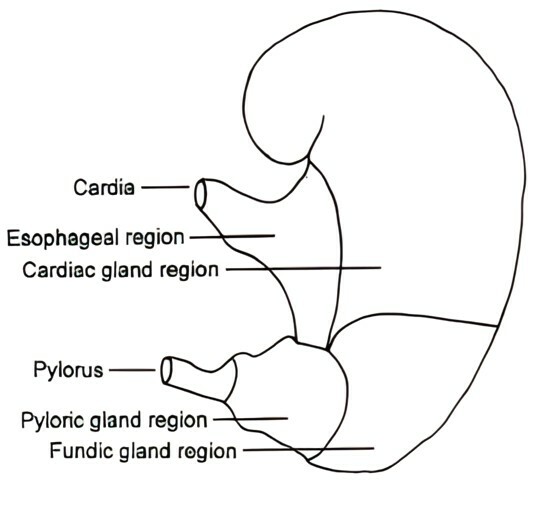
The stomach has four distinct areas which include the oesophageal, cardiac, fundic and pyloric regions (Figure 2). The oesophageal region is located at the entrance of the stomach from the oesophagus. This region of the stomach does not secrete digestive enzymes but has significance in that this is where ulcer formation in pigs occurs. Irritation in this area due to fine particle size, stress or other environmental factors can contribute to ulcer formation in swine. Once food passes though this region, it enters the cardiac region.
In the cardiac portion of the stomach, mucus is secreted and mixed with the digested food. Food then passes into the fundic region which is the first major portion of the stomach that begins the digestive process. In this region, gastric glands secrete hydrochloric acid, resulting in a low pH of 1.5 to 2.5. This reduced pH kills bacteria ingested with the feed. Other secretions in this region are present in the form of digestive enzymes, specifically pepsinogen. Pepsinogen is then broken down by the hydrochloric acid to form pepsin, which is involved with the breakdown of proteins.
Finally the digesta moves to the bottom of the stomach, which is the pyloric region. This region is responsible for secreting mucus to line the digestive membranes to prevent damage from the low pH digesta as it passes to the small intestine. The phloric sphincter regulates the amount of chyme (digesta) that passes into the small intestine. This is an important function not to overload the small intestine with chyme so proper and efficient digestion and absorption of nutrients occurs. In addition, once the chyme leaves the stomach, the material is quite fluid in consistency.
Small Intestine, Pancreas and Liver
The small intestine is the major site of nutrient absorption, and is divided into three sections. The first section is the duodenum. The duodenum is approximately 12 inches long and is the portion of the small intestine that ducts from the pancreas and the liver (gall bladder). The pancreas is involved with both exocrine and endocrine excretions. This means the pancreas is responsible for secretion of insulin and glucagon in response to high or low glucose levels in the body. In addition, it has exocrine functions of secreting digestive enzymes and sodium bicarbonate.
The digestive enzymes secreted break down (hydrolyse) proteins, fats, and carbohydrates in the chyme. In addition, the sodium bicarbonate serves a vital role to provide alkalinity so chyme can be transported though the small intestine without causing cell damage because of the low pH after leaving the stomach. The pancreas serves as the most vial organ in the digestive process for producing and secreting enzymes needed for the digestion of chyme and the prevention of cell damage due to pH.
In addition to the pancreas secreting into the duodenum, bile, which is stored in the gall bladder and produced by the liver, is secreted as well. Bile salts, which are the active portion of bile in the digestion process, primarily assist in the digestion and absorption of fat but also help with absorption of fat-soluble vitamins and aids pancreatic lipase in the small intestine. Finally, bile salts are necessary for the absorption of cholesterol, which takes place in the lower small intestine and are circulated to the liver via the portal vein.
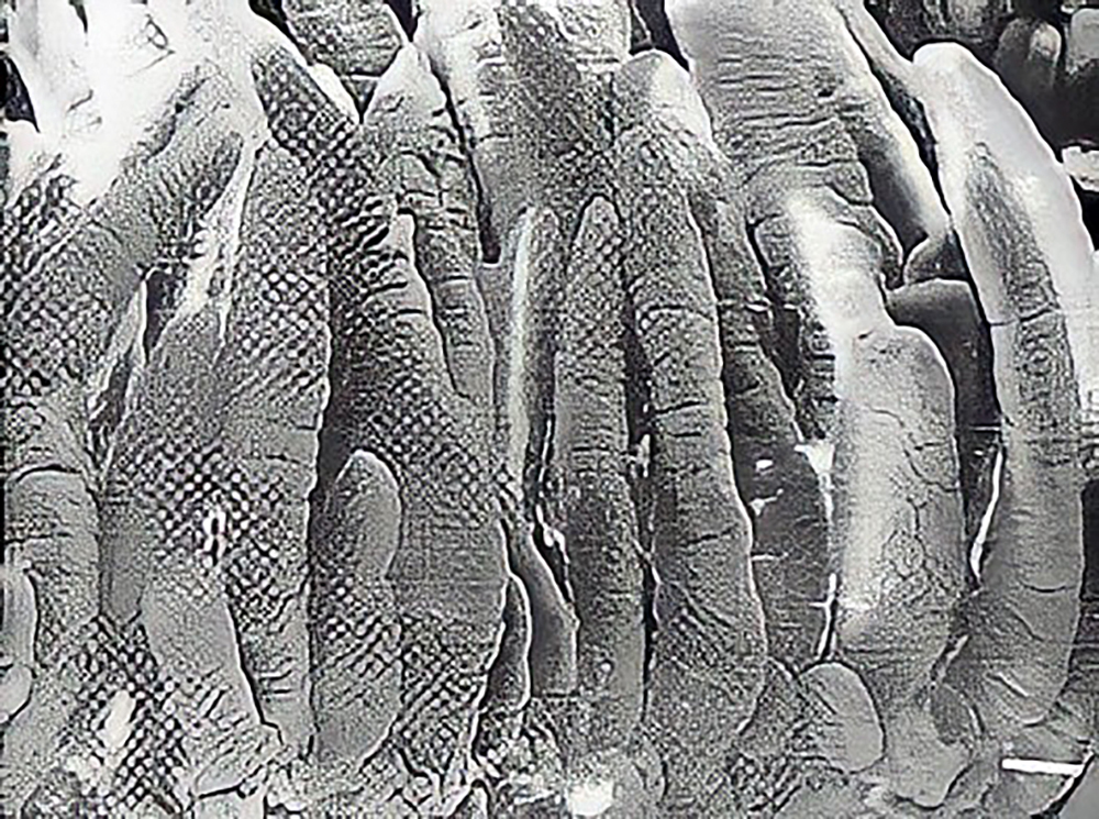
Once the chyme passes though the duodenum, the digestion process is in full swing. Upon leaving the duodenum, enters the middle portion of the small intestine, the jejunum. This portion of the small intestine involves both the further breakdown of nutrients as well as the beginning of absorption of nutrients. Nutrient absorption continues into the final section of the small intestine, the ileum. Absorption of nutrients in the jejunum and the ileum occurs in the area termed ‘brush border’, or the intestinal mucosa (Figure 3). The mucosa is comprised of finger-like projection called villi, which in turn contain more micro-size projections called microvilli. The tips of the microvilli form web-type structures called glycocalyx.
Amino acids and simple sugars released into the brush border membrane are absorbed into the microvilli first, then into the villi, and then pass into the circulatory system. Absorbed amino acids and simple sugars are taken directly to the liver via the portal vein. For dietary fat that is broken down and absorbed into the brush border, they enter the lymphatic system and are released into general circulation via the thoracic duct.
Large Intestine
The large intestine or hindgut encompasses four main sections. First, digesta from the small intestine passes into the caecum. The caecum has two sections, first a section that has a blind end, where material can not pass though. The caecum has a second portion where it connects to the colon, where digesta is passed to the rectum and anus where the remaining digesta is excreted.
The main function of the large intestine is the absorption of water. The chyme that passes through the small intestine and into the large intestine initially is very fluid. The large intestine epithelium has a large capacity for water absorption.
Once digesta passes though the ileum into the large intestine, no enzymatic digestion occurs. However, limited microbial enzymes activity does occur in the large intestine, which forms VFAs (volatile fatty acids). These can be readily absorbed in the large intestine. Generally these provide only enough energy to assist in the nutrient requirements of the epithelium of the large intestine. Also, B-vitamins are synthesised in the large intestine and are absorbed in a very limited amount, but not significant to alter nutritional supplementation of them.
With the majority of water removed, the digesta is condensed into a semi-solid material and is passed out of the rectum and anus.
Further Reading
You can view other papers presented at Swine Profitability Conference 2009 by clicking here.
June 2009