Bacteria
 9 November 2018
 4 minute read
Grouped under this heading are chlamydia, mycoplasma and other bacteria generally.
Chlamydia
These and anaplasma used to be classified separately from bacteria but are now regarded as bacteria. Chlamydia are the first of the very small bacteria that live inside or on the surface of a host cell and they like viruses are obligatory parasites. They are relatively unimportant in the pig but can be associated with respiratory disease, heart sac infection and in the case of Eperythrozoon suis, anaemia and infertility. They are also associated with jaundice and poor growth although the bacteria can also be commensal parasites, in which case they cause little if any problems. Chlamydia can also cause abortion and may be responsible for a new emerging disease. In the pig they are mostly respiratory spread or in the case of Eperythrozoon suis by biting insects or inoculation. Eperythrozoonosis can be diagnosed from blood smears examined under the microscope or more recently by PCRs.
Mycoplasma
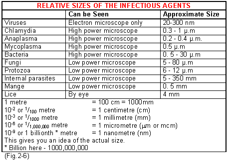
Mycoplasma are very tiny organisms less than half the size of other small bacteria. (See Fig.2-6). They can only just be seen under a high power light microscope. They tend to live and multiply close to the surface of cells. They are respiratory spread and are mainly responsible for enzootic pneumonia (Mycoplasma hyopneumoniae), and mycoplasmal arthritis associated with joint infections (Mycoplasma hyosynoviae).
Mycoplasma can be grown on solid media; however, this is not easy and can only be done reliably in a few diagnostic laboratories. It is time consuming and often the identification of one strain is complicated by the presence of other strains. Few laboratories are able to identify them in smears under the microscope but PCRs are now becoming available.
Mycoplasma hyopneumoniae is found in the respiratory tract of pigs and survives for only a few hours outside the pig. New herds can be established free of this particular organism. Then the main method of reintroduction of the disease is through the purchased carrier pig or by windborne spread from a nearby herd or a vehicle carrying pigs. Two other less important mycoplasma are Mycoplasma hyorhinis and Mycoplasma flocculare. The former can affect the smooth membranes that cover the joints causing lameness, arthritis and pericarditis (inflammation of the heart sac). Mycoplasma flocculare causes small lesions that may be mistaken for enzootic pneumonia. It is often involved in pneumonia complexes.
Other Bacteria
These can be readily seen under the microscope, particularly when they are stained. They are recognised by family group according to their shape, size, biochemical characteristics, antigenic characteristics and recently by identification of their DNA.
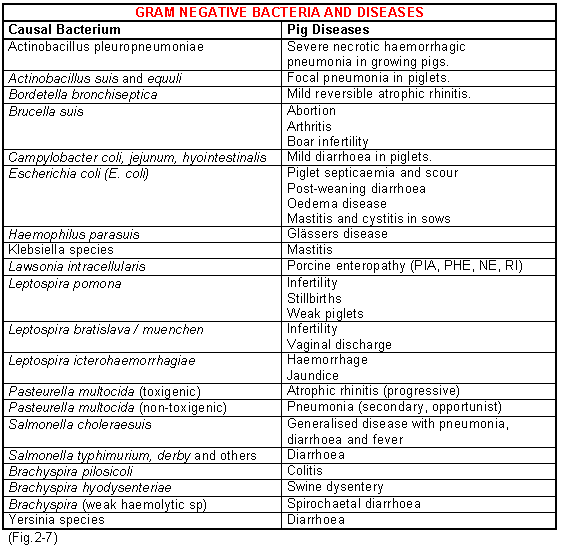
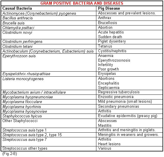
Most pathogenic bacteria can be grown easily in nutrient liquids and on solid media containing nutrients set in agar gel. Most grow profusely within 24 to 48 hours, although some such as the tuberculosis bacillus can take up to 3 weeks or more. They form little colonies consisting of billions of organisms and the shape and colour of these may be characteristic for a particular family or specific bacteria. A tiny smear from a colony spread on a glass slide and stained will give both the shape and the staining reaction of that particular bacteria. The stain commonly used is called a gram stain, and bacteria either stain positive (purple), or negative (red). This stain is of great help in carrying out primary observations. For example, the bacterium that causes meningitis in pigs, called Streptococcus suis type 2 is a small round organism in pairs or short chains that always stains positive or purple. Figs.2-7 and 2-8 show the different gram +ve and gram -ve bacteria and the diseases they cause.
Each bacterium has a number of specific characteristics that are peculiar to itself. Some, for example Anthrax bacillus, form spores which can survive outside the pig for many years. Organisms such as E. coli and salmonella can remain viable outside the pig for up to six months but Mycoplasma hyopneumoniae, the cause of enzootic pneumonia, probably for no longer than a few hours. Like viruses the survival of bacteria is also dependent upon the material surrounding them (faeces, soil, pus, urine, blood etc.), temperature, moisture and exposure to ultra-violet light. Again like viruses, they can survive indefinitely when frozen and for long periods in cold damp dark weather but only for a short time in very sunny weather. Such knowledge is important in controlling the spread of the disease.
Bacteria like viruses may attack specific parts of the anatomy or an individual system. For example, Actinobacillus pleuropneumoniae infects the pleura (smooth surface covering the lungs) and the lung tissue beneath. E. coli or salmonella invade the small intestine setting up an enteritis but Salmonella choleraesuis, the host adapted serotype of pig salmonellae, can affect the whole of the body including the lungs causing pneumonia.
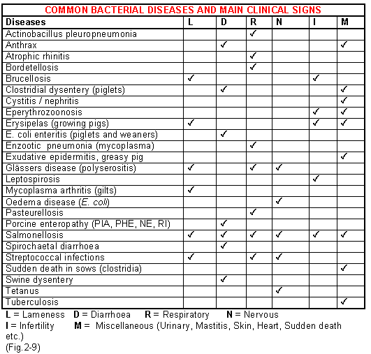
Most bacterial diseases are characterised by specific clinical signs and these are shown in Fig.2-9. The common routes of bacterial spread are by direct contact. close respiratory droplet contact, infected faeces or mechanical transfer on shovels, boots, vehicles etc. You will see in Fig.2-17 some of the distances recorded in the field when disease did not transfer between one farm and another. They can be surprisingly small. For depopulation and re-population purposes however, those diseases with an airborne transmission of less than 230m should, in practice, be extended to at least 900m.