Viruses
 9 November 2018
 5 minute read
What are viruses?
Pathogenic organisms that infect pigs can be listed in order of decreasing size and complexity and viruses are the smallest of the infectious agents. They cannot be seen by the type of light microscope that is used for looking at bacteria. They can only be seen through an electron microscope. Fig.2-0a.

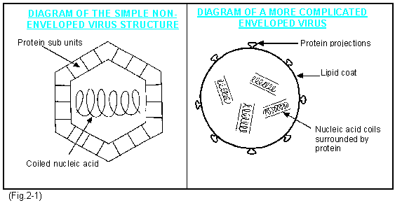
At their simplest they consist only of nucleic acid (i.e. their genes) and proteins which are arranged around the nucleic acid in a geometrical design and which protect the gene. (Fig.2-1). Some larger ones also have a loose outer coat (the envelope) which may contain lipids (fats) and carbohydrates which are derived from the hosts cell. They also contain proteins.
Viruses contain a genetic code in the nucleic acid but they do not contain the full mechanisms for their own multiplication. They have no energy-generating systems and lack chemicals such as enzymes required for their own reproduction. They depend entirely on those of the host.
This is the first important point to be aware of. Outside the host viruses are inert. They have no metabolic activity. Inside the host's cell they behave like pirates. Their genes using the host's nucleus take over control of part or all of the cells mechanisms. They code these mechanisms to make many more viruses exactly like the ones that invaded.. This activity usually damages or destroys the cells.
How do they get into cells?
The proteins on their surface stick specifically to receptors on the cells' surfaces. The cells then engulf them as if they were taking in particles of food but the particles are destructive invaders. They quickly loose their outer envelopes and the proteins covering their genes, which then take over from the cells nucleus and direct the cell to make more viruses. Some viruses (RNA viruses) replicate in the cytoplasm and some (DNA viruses) in the nucleus
The classification of a virus
All animal, plant and bacterial genes are made of a nucleic acid called DNA (deoxyribonucleic acid) which is found in the central nucleus of the cells and a further one RNA (ribonucleic acid) found in the cytoplasm. Virus genes however only contain one or the other.
The first broad classification is therefore into DNA viruses and RNA viruses. Fig.2-2 and Fig.2-3. These are then classified into families based on their shape, size and structure. N.B. No prion diseases (equivalent to BSE in cattle) are known to occur in pigs.
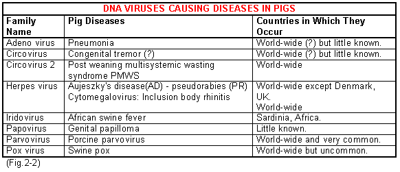
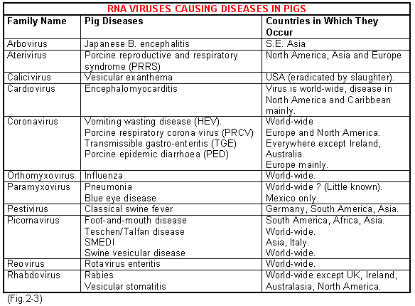
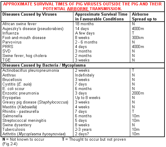
Fig.2-4 illustrates the approximate survival times of viruses that cause pig diseases. Some are extremely fragile and survive only a matter of days whereas others can persist for months. The survival times given are only approximate because in any virus they vary widely depending on the material it is on (faeces, salvia, blood, water, dust etc.), the temperature, the humidity, sunlight, and relative acidity. In general viruses survive long periods if frozen, fairly long periods in damp overcast cold weather, but only short periods in hot sunny dry weather. This is why virus diseases are more common in winter than summer. This information clearly is important in their control. Most but not all viruses are destroyed by strong acid or alkaline solutions and those that have a fat coating are quickly inactivated by fat solvents.
Some viruses can multiply sub-clinically in a pig for long periods of time (a carrier state) as in the case of aujeszky's (pseudorabies) disease and the cytomegalovirus that causes inclusion body rhinitis. Others can be carried sub-clinically for intermediate periods of 2 to 3 months such as PRRS but others such as TGE are usually eliminated after a week or two. However they may also persist in herds for long periods by successions of pigs post-weaning maintaining the infection.
Virus may be shed in saliva, urine, faeces, milk or in expired air from the lungs, or from vesicles on the skin.
The transmission may be by direct pig to pig contact, or indirect contact on farm machinery, pig trucks, or by vectors such as the wind, birds, flies, or discarded human pig meat products.
Clinical signs of disease
Viruses such as classical swine fever disseminate throughout the body and damage different organs thus producing a variety of clinical signs. Some such as TGE virus are more specific and disseminate sub-clinically initially to become concentrated in one or two target organs, which in this disease is the intestinal lining, causing diarrhoea. Some viruses can only multiply in one organ and swine influenza for example is generally only found in the lungs and respiratory tract.
Common pig viruses and their associated symptoms are shown in Fig.2-5.
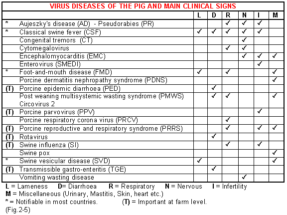
The diseases marked by an asterisk (*) are the major ones that are notifiable in many countries and controlled by eradication policies. The diseases marked by a T are the most important at farm level.
Diagnosis of virus infections
It is more difficult and expensive to grow viruses than bacteria in the laboratory. They have to be grown in living cell-cultures artificially in tubes or bottles or in the living cells of embryos in hens eggs. Some cannot be grown by either of these methods. So until fairly recently virus diseases were diagnosed by their clinical signs and post-mortem lesions and confirmed by serology. Serology requires two blood samples to be taken from each animal 1 to 2 weeks apart so that rising antibody levels can be demonstrated., A single sample is less helpful because you do not know whether the antibody levels are rising from a recent infection or are persisting from an old infection.
Over recent years new techniques have speeded up and simplified laboratory diagnosis because the virus does not have to be cultured. The use of the electron microscope directly on to faeces or samples from lesions has assisted in diagnosis, further aided by the fluorescent antibody test (FAT) that again can be carried out directly on faeces or lesion samples. The ELISA test is now commonly used in many diseases (e.g. Foot-and-mouth disease) to provide a rapid accurate diagnosis. New techniques such as polymerise chain reactions (PCRs) have been developed to demonstrate the virus genetic material (i.e. the DNA or RNA) in samples, and such tests for PRRS virus are now available. Their advantage is that they can accurately detect tiny amounts of the DNA or RNA. Unfortunately they are expensive if done in small numbers.
Treatment of a virus
- Some new medicines are available for the treatment of a few viral infections in human beings but they are expensive and not available for use in pig diseases. Since viruses have no cell wall and no metabolism of their own antibiotics will not destroy them, although they may help by preventing secondary bacterial infection. Hyper-immune antiserum might be helpful in some virus infections if given by injection, but in general they are not commercially available.