

Enzootic Pneumonia or Mycoplasma hyopneumoniae infection
Background and history
Enzootic pneumonia is caused by a tiny organism Mycoplasma hyopneumoniae. It is widespread in pig populations and endemic in most herds throughout the world. It is transmitted either through the movement of the carrier pigs or by wind-borne infection for up to 2 1/2 - 3km (1 1/2- 2 miles) if the climatic conditions are right.
The organism dies out quickly outside the pig, particularly when dried. It can however be maintained in moist cool conditions for two to three days. It has a long incubation period of two to eight weeks before clinical symptoms are seen. As an uncomplicated infection in well-housed and managed pigs it is a relatively unimportant disease and has only mild effects on the pig.
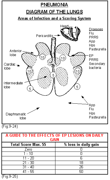
However if there are other infections present particularly App, Hps, Pasteurella, PRRS or SI, the pneumonia can become more complex with serious effects on the pig. Fig.9-24 shows the basic structure of the lobes of the lungs. EP always attacks the lower shaded areas of each of these lobes (anterior, cardiac, intermediate and anterior diaphramatic) causing consolidation of the tissues.
The extent of this consolidation in each lobe is scored out of either 5 or 10 depending upon the lobe affected. Thus a severely affected pig with all lobes involved would score 55. Occasionally, particularly in disease breakdowns the diaphramatic lobes will be involved as well. If more than 15% of lungs are affected it is highly probable that EP is present in the population. Herds that do not carry M. hyopneumoniae rarely show consolidated lesions of more than 1 to 2% and even then they are very small.
This scoring system can be used to assess the severity of disease and its effects on the pig. (Fig.9-25).
If EP is not present in the growing population then the effects of the other respiratory pathogens are very greatly reduced. It is therefore considered a prime organism that opens up the lung to other infections.
 |
Clinical signs
Acute disease
This is normally only seen when EP is introduced into the herd for the first time. For a period of six to eight weeks after entry there may be severe acute pneumonia, coughing, respiratory distress, fever and high mortality across all ages of stock. This picture however is extremely variable and breakdowns are experienced when disease is mild or inapparent.
Chronic disease
This is the normal picture where the organism has been present in the herd for some considerable time. Maternal antibody is passed via colostrum to the piglets and it disappears from seven to twelve weeks of age after which clinical signs start to appear. A prolonged non-productive cough, at least seven to eight coughs per episode, is a common sign around this time, with some pigs breathing heavily ("thumps") and showing signs of pneumonia. 30 to 70% of pigs will have lung lesions at slaughter.
Diagnosis
In most cases this is based on the clinical picture and examination of the lungs of pigs at post-mortem or at slaughter, combined possibly with histology
of the lesions.
 Lungs with gross Enzootic pneumonia |
However, these do not provide a specific diagnosis and in the herds supplying breeding stock or in special cases (e.g. litigation) it may be necessary to confirm the diagnosis by carrying out one or more of the following tests: ELISA tests, serum tests for specific antibodies, microscopic examination of stained touch preparations (TPs) of the cut surface of the lungs, fluorescent antibody tests (FATs), polymerase chain reaction (PCRs) tests and finally culture and identification of Mycoplasmal hyopneumoniae.
These tests are now widely available and many diagnostic laboratories cannot do them. The PCR is probably the most sensitive. FAT, serology and cultures are used in Denmark, but only FATs are available in some countries.
Similar diseases
Consolidation of the anterior lobes of the lungs at a low level can be caused by other respiratory pathogens including SI, PRRS, Hps certain viruses and other mycoplasma. Laboratory tests are required to differentiate them. Furthermore, all or some of these may occur as mixed infections together with Mycoplasma hyopneumoniae.
Causes
Enzootic pneumonia is caused by a tiny organismMycoplasma hyopneumoniae. It is widespread in pig populations and endemic in most herds throughout the world. It is transmitted either through the movement of the carrier pigs or by wind-borne infection for up to 2 1/2 - 3km (1 1/2- 2 miles) if the climatic conditions are right.
Prevention
The EP free breeding herd
- Most breeding organisations can supply breeding stock that is deemed to be EP free and so a new pig farm can be set up or an old one repopulated EP free. The question is whether it will remain so. One rule of thumb is that if you have an uninterrupted view of an infected EP herd, particularly if it is less than 3km (2miles) away, there is a definite chance that sooner or later your herd will become contaminated by wind-borne infection. If the herd you can see is a grower/finisher as distinct from a weaner producer than the risk is greater. The risk also increases the larger the infected herd. In a pig dense regions such as in the pig rearing areas of Eastern England, Germany, the Netherlands, Belgium, Eastern Spain, Quebec or Japan, it is impossible to remain free. You are likely to remain EP free indefinitely if your EP free herd is in a region of low pig density, such as parts of North or Southwest France, Northern Spain or the West USA. Likewise if the land is hilly or mountainous or on a sea coast.
To maintain an EP free breeding herd
- Keep it closed. Introduce genes only by AI, embryo transfer, or hysterectomy and fostering.
- Purchase only EP free stock from a reputable seed stock supplier, if possible from the same source herd every time, or at least from the same breeding pyramid. (It would be sensible to check whether the source herd is also free from lesions of App).
- Isolate incoming pigs for eight weeks and check that the EP free status of the donor herd has been maintained before moving them into your herd. (This is good practice whether your herd is EP free or not).
- To be ultra careful, mix sentinel pigs from your herd in with the new pigs in isolation, one to two weeks after their delivery. The sentinels can be slaughtered and their lungs examined before the pigs move in or they can be blood tested on entry to the isolation and again five weeks later. (Whatever the health status of your herd, EP free or not, it is a good practice to put pigs from your herd alongside the isolated gilts. This helps them to gradually adapt to the health status of your herd).
To maintain an EP free grower/finisher herd
- Purchase only from an EP free source but instead of an isolation period implement an all-in all-out policy, by site if possible, or if not by building.
- Again the location is paramount.
The herd with endemic EP
- Purchase stock with EP but, depending on the health status of the herd, make sure the pigs are free from swine dysentery, mange, App and PRRS.
- Carry out isolation and monitoring procedures as above. Six weeks instead of eight weeks is probably enough. If your herd has a low health status and is intensively housed it may be helpful to medicate the feed for the incoming stock if they are EP free. Use lincomycin, tiamulin or tilmicosin for the last two weeks in isolation, or the first four weeks in your herd. This allows them to become immune without becoming ill. Discuss with your veterinarian.
- Keep a broad parity spread in your sows. Sows of second parity onwards are more immune than first litter gilts and pass a better immunity to their piglets.
- Check the faeces of the growing pigs for ascarids and if present keep them under control by routine worming and all-in all-out housing procedures.
- If you are having clinical problems and poor growth later in growing pigs vaccinate young piglets against EP as per manufacturers instructions.
Increased disease is associated with the following (consider changes):
- Overcrowding and group sizes in any one environment of more than 200.
- Variable temperatures and poor insulation.
- Variable wind speeds and chilling.
- Low temperature, low humidity environments.
- Houses with poor hygiene and high levels of carbon dioxide and ammonia.
- High dust/bacteria levels in the air.
- Pig movement, stress and mixing.
- A shortage of trough space.
- Housing with a continuous throughput of pigs.
- Other concurrent diseases.
- Poor nutrition.
- Dietary changes at susceptible times.
- Slatted floors and liquid waste.
- Less than 3 cu.m. air space/pig and 0.7 sq.m. floor space/ pig.
- Houses that are too wide for good air flow control.
- Presence of PRRS.
- Presence of aujeszky's disease.
- Presence of App.
- Presence of swine influenza.
- Purchasing from different sources.
Therefore to keep EP and respiratory disease under control:
- Optimise stocking levels by pen and house. This will not only reduce clinical signs but the energy that had been required for immunity will be available for growth and result in faster throughput. Thus the number of pigs sold per year can remain the same as at higher stocking levels.
- Optimise the ventilation and improve the hygiene to reduce noxious gases.
- Improve the insulation if necessary and maintain constant temperature control.
- Reduce the dust and bacteria in the air by changing to wet feed or to less dusty dry feed. (Note increasing ventilation does not decrease dust and may increase it).
- Check the nutrition and time of feed changes.
- Organise the grower/finisher stages so that moving and mixing is minimal.
- Operate an all-in all-out system wherever possible.
- Vaccination. Highly efficient EP vaccines have recently come to the market that reduce lung lesions by up to 95%. Some can be applied at one and three weeks of age and this provides excellent control during the growing and susceptible period. Vaccine could also be applied to EP negative herds if considered at risk. The use of enzootic pneumonia vaccines in herds with complex respiratory problems has revolutionised respiratory disease control particularly if used in conjunction with segregated weaning and segregated disease control techniques - the latter on combined breeding/finishing operations.
If you have a problem on your farm consider the following simple criteria in making the decision to vaccinate:
- The presence of Mycoplasma hyopneumoniae.
- A continual level of respiratory disease.
- Primary or secondary infections associated with PRRS, influenza, pseudorabies and Actinobacillus pleuropneumoniae.
- Heavy bacterial challenge.
- The necessity for continual in-feed medication.
- Variable and poor growth associated with respiratory disease.
- Weaning to slaughter mortalities of more than 4%.
- Finally, the cost of vaccination should be equal to or less than costs of the potential reduction in mortality and in-feed medication. Improvements in daily gain and feed efficiency become the bonus. In severely affected herds a cost benefit ratio of up to 5:1 has been achieved.
- Field experiences indicate that all herds should vaccinate growing pigs from one week of age onwards.
Treatment
In the herds in which the disease has become endemic a decision to medicate feed should be based on the following:
- Variable growth in pigs from 10 to 20 weeks of age.
- Ongoing pneumonia treatment of individuals at more than 2.5% of the pig population.
- Of lungs scoring more than 15.
- Active lesions - raised above the level of the lung surface and moist or wet.
- Methods of medication have been extensively dealt with in chapter 4 page.
Acute disease (Herd breakdown) - Consider the following:
- Medicate pigs between weaning and 16 weeks of age for 4 to 8 weeks with 500g/tonne of CTC or OTC and then reduce this to 200-300g/tonne. Alternatively 200g to 400g tilmicosin per tonne for 15 days or tiamulin 100g per tonne for 14 days.
- Inject severely affected individual pigs with either long-acting OTC, tiamulin, lincomycin, valnemulin, tilmicosin or penicillin/streptomycin.
- If pigs become affected soon after weaning inject with OTC LA at weaning time or one week prior to the onset of disease.
Chronic disease
- Identify the point at which disease is occurring and apply strategic medication, either in feed, in water or by injection using the medicines outlined above.
- For strategic medication use tetracyclines 500-800g/tonne, 220g/tonne of lincomycin or 100g/tonne of tiamulin and feed for 7 to 10 days or 200g - 400g / tonne tilmicosin for up to 10 - 15 days commencing one to three weeks prior to the anticipated time of the disease starting.


