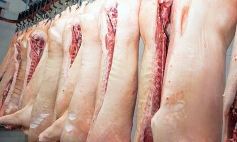


VLA: Congenital Hyperostosis in Newborn Piglets
UK - Congenital hyperostosis has been observed in piglets, according to the Veterinary Laboratories Agency's (VLA) Monthly Scanning Surveillance Report for February 2011.Alimentary tract diseases
Clostridium perfringens Type C

Clostridium perfringens type C was confirmed as the cause of enteritis in a case involving piglets in an outdoor rearing unit which had developed diarrhoea within the first few days of life. Most appeared to respond to antibiotic medication if caught early although there was a significant ongoing mortality. Necropsy of an affected piglet revealed severe emphysematous change to the serosal surface of the ileum with many gas bubbles present in the subserosal tissue. Much of the small intestine was also heavily congested, the content was liquid and blood tinged. Clostridium perfringens Type C infection was confirmed by the demonstration of both alpha and beta toxin in gut content.
Respiratory Diseases
Influenza
Thirsk diagnosed swine influenza as the cause of sudden onset lethargy and respiratory distress in a group of 180 eight-week-old pigs, with 100 per cent morbidity and less than 1 per cent mortality. Although Influenza A RNA was detected, the strain involved was not pandemic H1N1 (2009).
Pasteurellosis
Two recently farrowed gilts with a history of respiratory signs, milk drop and weight loss, were submitted to Sutton Bonnington for post-mortem examination. The pathology was similar in both gilts with a bronchopneumonia with dark purple areas of consolidation with a rubbery consistency, typical of enzootic pneumonia. There was mucopurulent exudate in the trachea with septicaemic-type lesions. P. multocida was isolated from the lung and M. hyorhinis and M. hyopneumoniae were detected and identified from lung tissue of both animals. Histopathology confirmed a subacute to chronic lymphoplasmacytic bronchointerstitial pneumonia with purulent bronchiolitis and acute multifocal exudative pneumonia, likely associated with P. multocida invasion. Neither PRRS nor Influenza virus were detected by PCR.
Other diseases
Four sudden deaths of 18 to 28-week-old finishers occurred over a 48-hour period on an indoor breeder-finisher farrowing weekly. Apart from low level coughing in growing pigs, herd health was good. Two dead pigs were submitted to Bury with gross lesions suggestive of a septicaemia; there was a cooked meat appearance to the musculature, reddening of the ventral skin and fibrin stranding in the peritoneal cavities. One pig had a severe fibrinous and pericarditis. Streptococcus suis 2 was detected in meningeal smears from both pigs and the same organism was isolated from the meninges and pericardium, consistent with streptococcal septicaemia. There was no clear predisposing factor for the upsurge of streptococcal disease late in rear; no involvement of PRRSV or swine influenza virus was detected.
Congenital hyperostosis of newborn pigs

Thirsk diagnosed Congenital hyperostosis of newborn pigs as the cause of musculoskeletal abnormalities which had reportedly affected a total of 16 piglets on a 600 sow indoor, fortnightly batch farrowing, farrow to finish unit. Six litters had been affected, typically containing one to four affected piglets. One affected piglet was submitted for necropsy which revealed marked thickening of the radius and ulna of both forelimbs, with tight shiny skin (see Figure 2). Congenital hyperostosis is described in the literature and is suspected to be a rare autosomal recessive condition which is similar to Caffey’s disease in humans, where the mode of inheritance and affected gene has been determined. Plans are ongoing to monitor the situation and collect further affected and unaffected piglets to gather an archive of tissue which may be used in the future for genetic testing by the breeding company.
Neurological Diseases
Bury necropsied a gilt from a recently farrowed batch of 112 sows and gilts on an outdoor herd, which had died suddenly. The gilt had farrowed 11 live pigs 10 days earlier and was found recumbent outside her hut on the morning of submission and rapidly died. The gilt was in good body condition and milking well and there were no specific gross findings to indicate the cause of death. Histopathology revealed polioencephalomalacia concentrated within the laminar cortex with a mild mixed perivascular reaction including significant numbers of eosinophils. These changes were highly suggestive of early salt poisoning/water deprivation syndrome. Subsequent investigation failed to identify any problems with the available water supply.
Further Reading
|
| - | Find out more information on the diseases mentioned by clicking here. |
| - | You can view the full report by clicking here. |


