AHVLA: PCV2-associated Disease on Newly-established Unit
UK - Intestinal torsion on an outdoor unit, outbreaks of Glässers disease in the early post-weaning period and porcine circovirus-2-associated disease on a newly-established unit have been noted in the AHVLA's pig disease surveillance for March 2013. 12 June 2013
 7 minute read
Alimentary Disease
Gastric ulceration causing losses on two units
A problem of anaemia and weight loss in pigs from 14-15 weeks old was first observed on a 2100 pig nursery-finisher unit in January 2013 and then investigated in March. Approximately 10 pigs were affected in a group of 250 and all affected pigs died. The problem subsided but after two more pigs became pale, one 18-week-old pig was submitted to Sutton Bonington for investigation. A perforated gastric ulcer at the pars oesophagus had resulted in a fatal peritonitis. It was suggested that risk factors for gastric ulceration be examined, including food particle size in the pelleted diet.
On an unrelated unit, a six-month-old maiden gilt was submitted to Winchester with a history of malaise and salivation prior to being found dead on an outdoor unit. The carcase was pale but in good bodily condition. A large gastric ulcer was present with evidence of historic and recent haemorrhage. The distal oesophagus was also thickened due to hyperkerat osis, likely due to irritation from reflux of gastric contents.
Intestinal torsion on an outdoor unit
The carcase of an adult sow was submitted to investigate sudden death of five sows from a group of 120 on an outdoor unit. Post mortem examination revealed a mesenteric torsion affecting the small intestine; intestinal loops were gas and fluid-filled and markedly dilated. Intestinal torsions can be related to the consumption of large amounts of stones or sand which accumulate within the lower small intestine and caecum. Groups of replacement gilts are someti mes prone to ingest excessive soil when they are introduced to outdoor conditions. Management issues such as irregular feeding or excitement and excessive activity around feeding may also predispose to these dramatic intestinal torsions.
Weight loss and diarrhoea in sucking pigs due to coccidiosis and rotaviral enteritis
Three live ten-day-old piglets were submitted to Thirsk to investigate diarrhoea affecting a few litters from both gilts and sows. The diarrhoea was typically pasty and also resulted in weight loss. Post mortem examination revealed voluminous yellow intestinal contents and mesenteric lymph node enlargement. Rotavirus was detected in the faeces of one of the three piglets and histopathological examination revealed severe villus atrophy and loss with marked crypt hyperplasia. Numerous coccidial forms were also seen within villus enterocytes. Coccidiosis with rotaviral enteritis was diagnosed as the cause of problem.
Respiratory Disease
Increased mortality in growers due to bacterial pneumonia
A fresh pluck was submitted from a pig found dead on a unit where approximately 20 per cent of 500 10-week- old pigs were coughing with sudden deaths of good pigs and an increase in postweaning mortality to 7 per cent over the previous two to three weeks. The unit was a continuous indoor nursery-finisher with weaners entering weekly from a single breeding source. A severe bronchointerstitial pneumonia was present affecting all lung lobes with a cranioventral distribution. Pasteurella multocida and Actinobacillus pleuropneumoniae serotype 3, 6, 8 were both isolated, together with Streptococcus suis type 2 from the lung. Mycoplasma hyopneumoniae was detected by DGGE; however histopathological findings in the lung were primarily of a suppurative pneumon ia associated with bacteria, making the Pasteurella multocida and Actinobacillus pleuropneumoniae likely to be of most clinical significance. No viral involvement was detected.
Outbreaks of Glässers disease in the early post-weaning period
Three dead five-week-old weaners were submitted to Bury St Edmunds from an indoor nursery-finisher on which weaners from one source were lame, lethargic and suffered 10 per cent mortality. Disease appeared to show a poor response to amoxicillin treatment. Post mortem examination revealed fibrinous polyserositis and cranioventral pneumonia as well as variable degrees of typhlocolitis with diarrhoea. Glässer’s disease was confirmed as the cause of the polyserositis by isolation of Haemophilus parasuis in spite of recent antimicrobial treatment, together with salmonella due to Salmonella Typhimurium phage type 193. No underlying viral disease was detected and the poor response to amoxicillin may have been due to the concurrent salmonellosis, as the salmonella isolated showed in-vitro resistance to amoxicillin. This is not an uncommon resistance in salmonella isolates from pigs.
Glässer’s disease was also diagnosed as the cause of respiratory disease of variable morbidity and approximately 2.5 per cent mortality which began soon after pigs arrived on an outdoor unit rearing in tents. The pigs were vaccinated for Mycoplasma hyopneumoniae, PCV2 and PRRS. One euthanased and one dead pig were submitted; in both there was a fibrinous polyserositis from which Haemophilus parasuis was isolated, confirming the diagnosis.
Systemic Disease
Porcine circovirus-2-associated disease on a newly-established unit
Six live pigs aged nine and 12 weeks old were submitted from a newly-established indoor farrow to finish PPRSv and Mycoplasma hyopneumoniae negative unit, to investigate two distinct clinical presentations - wasting and meningitis - affecting growing pigs. In the pigs with signs of meningitis, pigs showed fibrin stranding in the abdominal cavity and marked engorgement of meningeal blood vessels, while the wasting pigs had pneumonia, enlarged mediastinal, mesenteric and sublumbar lymph nodes and varying degrees of polyserositis. Haemophilus parasuis was isolated from the pneumonic lung of one of the wasting pigs. Histopathology confirmed severe neutrophilic purulent meningitis in the meningitis pigs, although no bacteria were isolated. It is possib le that the meningitis was also attributable to Haemophilus parasuis and that this organism was only cultured from the lung due to its fastidious nature. An autogenous vaccine for Haemophilus parasuis was already in use. Histopathology also confirmed significant convincing lymphoid lesi ons consistent with porcine circovirus 2 associated disease (PCVAD) in the lymph nodes and spleen from the wasting pigs and Brachyspira pilosicoli was isolated from the intestine of those with mild diarrhoea. Piglets were not vaccinated with a PCV-2 vaccine and advice to commence vaccination was given in the face of this PCV-2 clinical manifestation.
Mulberry heart disease causing mortality
Fixed heart tissue from eight and twelve-week-old pigs was submitted for histopathology to investigate an ongoing problem of increased mortality in growing pigs. Around 30 from a batch of 1,500 had died at the time of submission. The morphological diagnosis from histopathology was multifocal acute myocardial necrosis consistent with a diagnosis of Mulberry heart disease. Vitamin E injections were considered for the remainder of the group to try and limit the high level of mortality, and the need for dietary supplementation for following batches was to be investigated.
Streptococcus suis infection involved in sudden deaths
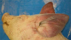
A vegetative valvular endocarditis and pericarditis causing heart failure was diagnosed as the cause of death of a 10-week-old pig submitted from an outdoor breeding unit on which four of 150 10 to 12-week- old pigs had died in the previous few days. Within the group, one tent was also affected with respiratory disease. Dying pigs were seen to have red-purple ears prior to death. In the submitted pig there was marked deep red discolouration of both ears, as illustrated in Figure 1, together with marked pulmonary oedema, both likely to be secondary to the heart failure and probable bacteraemia. An untypable Streptococcus suis was isolated from the valve lesion and no underlying viral involvement was detected. It was recommended that if respiratory disease and deaths continue, submission of further typical cases would be worthwhile to determine whether this was a sporadic finding and whether there was underlying viral disease in the group.
Endocarditis in group of growers dying suddenly
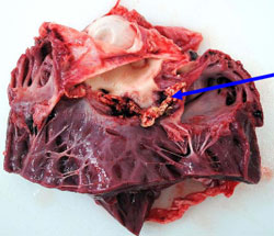
A 10-week-old grower pig was very lethargic and died in spite of veterinary treatment. This was the third pig out of nine to have died suddenly over a two-week period. Post mortem examination at the Royal Veterinary College/AHVLA surveillance centre revealed a marked serofibrinous peritoneal and pleural effusion. There were extensive fibrous adhesions between the epicardium and pericardial sac, and opening the heart revealed vegetative nodules on both semilunar and tricuspid valves (Figure 2). No significant bacteria were isolated, likely due to the recent antibiotic treatment. Endocarditis in pigs is most commonly associated with Erysipelothrix rhusiopathiae or Streptococcus suis. Submission of further cases, preferably untreated, was encouraged.
Miscellaneous Disease
Neoplasia of the adrenal gland causing fatal haemorrhage
The sudden death of a heavily pregnant adult sow following transportation was found to be due to an intra-abdominal bleed originating from the renal artery. The cause of the arterial rupture was infiltration of the tunica layers by a malignant sarcoma of the adrenal gland. The right adrenal gland and tumour was approximately 25cm in diameter and comprised whitish homogenous tissue interspersed with fine haemorrhages.
Visit our PMWS/PCVAD page by clicking here.