AHVLA Reports Finisher Deaths Due to Septicaemic Pasteurellosis in March
UK - The Animal Health and Veterinary Laboratories Agency (AHVLA) reports that during March 2014, finisher deaths due to septicaemic pasteurellosis was observed. Besides this, there was also an unusual outbreak of oedema disease causing ataxia and hindlimb weakness due to severe polyarthritis in finishers. 19 May 2014
 7 minute read
Respiratory Disease
Finisher deaths due to septicaemic pasteurellosis
Pasteurellosis was diagnosed by Bury St Edmunds when two fresh plucks were submitted to investigate widespread respiratory disease in 18-week-old pigs on an indoor finishing unit. Clinical signs were reported to have been ongoing for more than two weeks and the pigs had been treated with in-water antimicrobial. There was cranioventral pulmonary consolidation in both plucks and a fibrinous pericarditis. Pasteurella multocida was isolated from multiple sites. Histopathology was consistent with a bacterial pulmonary infection and there was no convincing histological evidence of either viral or mycoplasmal involvement; PCRs for swine influenza and PRRSV did not detect these viruses. There was also no evidence of PCV2-associated disease. As respiratory disease had been ongoing for several weeks prior to submission, earlier involvement of viruses, in particular swine influenza, could not be ruled out. Bloods were submitted from the cohort of pigs at the same time as the plucks and swine influenza serology pointed to previous exposure to pandemic H1N1 2009. The fibrinous pericarditis associated with Pasteurella multocida infection, and certain features of the histopathology, suggests that the pasteurellosis was septicaemic and would account for the deaths of the pigs.
Systemic Disease
Lack of milk suspected as cause of neonatal piglet deaths
Three two-day-old piglets were submitted to Preston to investigate why seven of 10 piglets from one litter had died within two days of birth. The piglets had completely empty gastro-intestinal tracts. There was no evidence of septicaemia and no bacterial pathogens were isolated by culture. It was suggested that the sow be examined for the presence of milk and checked for mastitis.
Wasting in weaned piglets
Six pigs, three dead and three live, were submitted to Preston to investigate a six-month history of wasting one to two weeks post weaning. The pigs were vaccinated against Mycoplasma hyopneumoniae and PCV2. Wasting pigs were isolated and treated with antimicrobials but the condition caused approximately one per cent mortality. Post-mortem examination of the first batch of pigs aged 8-9 weeks identified umbilical abscesses and one pig with hydronephrosis and hydroureter but no findings to account for the group problem. No gross lesions were identified in live 5-6 week-old pigs to account for the problem. PRRSV was not detected and histopathology was unremarkable. Overall there was no evidence of an infectious problem and advice was given to reassess the management practices and nutrition around the periweaning period.
Erysipelas in finisher in continuous management system
Swine erysipelas was diagnosed when a pig showing respiratory disease was euthanased and submitted. The pig came from a small indoor commercial breeder-finisher with an ongoing respiratory disease problem showing a recent upsurge and five deaths in the week prior to submission. The pig had a severe vegetative valvular endocarditis affecting the left atrioventricular valve and Erysipelothrix rhusiopathiae was isolated. No viral involvement was detected. The finisher accommodation was strawed solid floor pens managed on a continuous basis and it is possible that this led to a build up of environmental challenge with the organism over the winter period.
Chronic pericarditis resulting in breeding gilt death
Two of 200 seven-month-old gilts died within a week on an indoor breeding unit. The gilts were vaccinated for erysipelas and porcine parvovirus. The submitted gilt was in good body condition with non-specific findings (enlarged submandibular lymph nodes and fine fibrin stranding in the abdominal cavity) but also a severe widespread pericarditis which was chronic but active. The pericarditis was sufficiently severe to have compromised cardiac function and, as no other cause was identified following further testing, was considered to be the cause of death due to cardiac failure. The pericarditis probably reflected earlier bacterial disease, for example due to Haemophilus parasuis.
Massive testicular swelling due to a malignant tumour in breeding boar
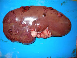
A single adult boar was reported to have been affected for approximately one month with swelling of one testicle, with no other clinical signs at this stage. In the four to five days prior to submission, the boar became increasingly lethargic and inappetent and had lost body condition. The boar was euthanased by barbiturate injection and submitted to Bury St Edmunds. The left testis was massively enlarged with a cream coloured solid homogenous mass replacing the head of the epididymis and extending around the testis. The right testis was not enlarged but had a single focal cream coloured lesion within its substance at the cranial pole, well demarcated from adjacent testicular tissue. Multiple similar lesions were present in liver, heart, kidney (Figure 1) and other lymph nodes. The gross findings were suggestive of neoplasia, affecting multiple organs, and histopathology confirmed multicentric lymphosarcoma.
Nervous Disease
Unusual outbreak of oedema disease causing ataxia
Oedema disease was diagnosed at Langford as the cause of nervous signs mainly consisting of ataxia, weakness and recumbency as well as malaise and anorexia. The small herd comprised three groups of pigs; 45 recently weaned, 29 weighing around 17kg, and 14 older pigs weighing between 70 and 130kg. The pigs were bought in from different units at the same time. At least 14 pigs across all three groups died over about three weeks. Post-mortem findings were non-specific with no typical oedema of the eyelids, gastric mucosa or mesocolon visible. Pulmonary oedema and increased peritoneal fluid with scant fibrin stranding was found in some of the six pigs submitted. Initially Streptoccous suis meningitis was suspected but no pathogens were isolated from the meninges. Histopathology supported the possibility of oedema disease when a fibrinoid vasculopathy was detected in brain and lymph node and E. coli serotype O139:K82 (strain E4) was isolated in pure culture from the small intestines of subsequent submissions confirming the cause of disease. Amongst the submissions was one pig with diarrhoea due to a different manifestation of the E4 E. coli infection. This pig had a haemorrhagic enteropathy and small intestinal thickening and corrugation not unlike porcine intestinal adenomatosis. E4 E coli was isolated in pure growth from the intestines.
This outbreak was unusual in that oedema was not evident before, or at, post-mortem examination, possibly because some pigs were euthanased and lesions had not fully developed. Oedema disease most commonly affects pigs one to four weeks after weaning. Here much older animals were also affected, which may have been due to later challenge following their transport and introduction into an environment contaminated with the oedema disease-producing strain of E. coli. Control can be difficult and centres on hygiene, controlling feed levels, high quality nutrition and sometimes a reduction in stocking rate.
Sapelovirus outbreak causing nervous disease
Unusual nervous signs and deaths in post-weaned pigs prompted the submission of, first, a fixed brain and then affected pigs to Thirsk from a 400-sow farrow to finish unit. This unit also had problems with Streptococcus suis meningitis which usually responded to antimicrobial treatment. However, prior to submission, one to two pigs from the same pen in each batch of 200 pigs weaned fortnightly presented with different nervous signs that did not respond to treatment. Pigs were affected approximately two weeks after weaning and were reported to be going off their front legs and walking backwards. They progressively worsened, showing nystagmus, then lateral recumbency but no paddling. Gross post mortem examination was unremarkable but brain histopathology revealed a severe nonsuppurative and necrotising polioencephalitis and panencephalitis. These findings were indicative of a neurotropic viral infection and were very similar to those in a recent outbreak of porcine sapelovirus in the Bury St Edmunds region. Immunohistochemistry and PCR for porcine sapelovirus confirmed this diagnosis and ruled out teschovirus involvement. Interestingly, this is the second outbreak of sapelovirus in 2014. The most recent case diagnosed by AHVLA prior to 2014 was in 2008. Affected pigs did not respond to nursing and it was advised that cases presenting with signs typical of the sapelovirus be culled promptly if they showed no response to antimicrobial treatment.
Musculo-skeletal Disease
Hindlimb weakness due to severe polyarthritis in finishers

Following the diagnosis of porcine sapelovirus earlier in rear on a nursery-finisher unit, several pigs around 14-weeks-old were reported to be showing hind limb weakness and reluctance to rise. Some of these had died and there had been a poor response to antimicrobial treatment. There was concern that this was a further manifestation of porcine sapelovirus. One typical case was euthanased and submitted to Bury St Edmunds. A severe fibrinosuppurative polyarthritis was present affecting all joints including the atlanto-occipital joint. No bacterial pathogens were isolated and Mycoplasma hyosynoviae was not detected by PCR. The pig had been treated prior to submission and this may have affected culture results; the detection of polyarthritis explained the clinical signs and ruled out the involvement of porcine sapelovirus.