APHA Report: PRRS, Bowel Oedema, Bacterial Joint Infections
UK - The Animal and Plant Health Agency (APHA) has reported incidents including respiratory disease involving porcine reproductive and respiratory syndrome (PRRS), sudden deaths from bowel oedema and bacterial joint infections with underlying PRRS in its Pig Disease Surveillance Monthly Report for December 2014. 17 April 2015
 9 minute read
 By:
By: Upward trend in porcine reproductive and respiratory syndrome diagnoses
Analysis of the seasonality of porcine reproductive and respiratory syndrome (PRRS) diagnoses in GB shows that the last quarter (October to December) of 2014 had the highest quarterly diagnostic rate for PRRS since 2004 when the PCR test was introduced, improving diagnosis.
Six percent of the APHA PRRS diagnoses were reproductive disease outbreaks. The remainder were all in post-weaned pigs.
Disease due to PRRS in post-weaned pigs is often associated with other pathogens or disease conditions, in part reflecting the immunosuppressive nature of the virus.
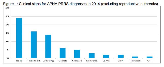
The three most common diagnoses made in submissions with PRRS at APHA in 2014 were Streptococcus suis, salmonellosis and pasteurellosis. This helps explain why clinical signs in PRRS outbreaks can be variable.
Figure 1 shows the clinical signs reported to APHA for outbreaks during 2014; respiratory signs, pigs found dead and wasting being the three most commonly described.
The fact that PRRS is often identified together with other pathogens emphasises the importance of full diagnostic investigation in disease outbreaks.
There is guidance on accessing diagnostic support and on sampling and testing available on this link: http://ahvla.defra.gov.uk/vet-gateway/surveillance/diagnostic-support.htm
Reproductive Diseases
Sporadic abortion with bacterial infection
Abortion due to an untypeable Escherichia coli was detected at Sutton Bonington following the submission of four foetuses and associated placenta from a gilt.
The gilt was one of 30 purchased in-pig and of high health status. The gilt was described as clinically normal in herself but had aborted 16 piglets on the evening prior to submission.
The placentas appeared unremarkable and gross findings in the foetuses were limited to increased volumes of serous fluid in abdominal and thoracic cavities. Bacteriology recovered pure growths of non-haemolytic Escherichia coli from stomach contents.
Screening for other infectious agents of porcine abortion did not identify another infectious. It was thought likely that this was a sporadic incident.
Enteric Diseases
Outbreaks of clostridial enteric disease in neonatal pigs
Two outbreaks of clostridial enterotoxaemia were diagnosed, both likely due to Clostridium perfringens type A and in two-day-old piglets submitted from outdoor units.
In the first case, watery yellow diarrhoea was reported to be affecting nearly half the litters with preweaning mortality increased to 20%. In the second case, two of 20 litters showed profuse watery diarrhoea affecting most piglets in these litters.
In both of the submissions, alpha toxin was detected in small intestinal contents of one piglet and histopathology revealed acute necrotic enteritis with large numbers of large rod-shaped bacterial in association with necrotic debris, supporting the diagnosis of a clostridial cause.
Histopathology was not suggestive of viral disease.
Unusual late occurrence of enteric colibacillosis
A rectal swab was submitted to investigate the sudden death of a single 20-week-old pig in good body condition from a group of 20 finishers on an indoor breeder-finisher unit.
On-farm post-mortem examination revealed a fluid-filled reddened intestinal tract and Escherichia coli strain E4 (serotype O139:K82) was isolated which is an enteropathogenic strain of E.coli associated with both enteric disease and oedema disease. No Salmonella species were isolated.
With the limited material submitted, it was not possible to investigate the significance of this isolate further, the post-mortem findings raise the possibility of an unusually late occurrence of enteric colibacillosis.
The same E. coli strain was isolated from an outbreak with a more typical clinical history for postweaning enteric colibacillosis.
Twenty of 500 four-week-old pigs which had just been weaned were showing light brown diarrhoea but no reported deaths.
Lawsonia intracellularis detected in diarrhoeic pigs
Diarrhoea and wasting of low morbidity was suspected to be due to Lawsonia intracellularis on a finishing unit of 1000 13-week-old pigs.
A faecal sample submitted was positive for L. intracellularis DNA supporting this suspicion and no Salmonella were isolated, however, it is preferable for pathology consistent with the disease to have been confirmed to make a diagnosis of disease due to L. intracellularis.
Salmonellosis diagnosed in pigs following likely Glässer’s disease
Pigs were submitted to Thirsk when eight-weeks-old to investigate diarrhoea starting about a week after weaning. The affected pigs wasted markedly despite treatment but did not die.
Post-mortem examination revealed all the pigs to have varying degrees of typhlocolitis with some also showing peritonitis, pleuritis and pericarditis. The only pathogen identified was Salmonella Typhimurium Copenhagen phage type U288.
The salmonella infection was consistent with the typhlocolitis and may have contributed to the peritonitis, pleuritis and pericarditis but it was suspected that Glässers disease, or another bacterial cause of polyserositis, was involved earlier.
In incidents with disease occurring in the early post-weaning period, submission of cases early in the course of disease is ideal.
In batch systems with only one age group present, this is not always possible if there are no younger pigs on the unit at the time a clinical problem of poor response to treatment is noted.
Some submitted pigs had been recently treated with amoxicillin, the Salmonella isolated was resistant to this antimicrobial, as are most Salmonella causing disease in pigs.
Respiratory Disease
Several incidents of respiratory disease involving porcine and reproductive syndrome
Porcine and reproductive syndrome (PRRS) virus was detected by PCR in pooled sera from 17-week-old indoor finishers which had been coughing for several weeks in the Bury St Edmunds region.
Fifty percent of the pigs were affected with low mortality. PRRS was also diagnosed at Bury St Edmunds in ten to 12-week-old pigs from an indoor breeder-finisher unit together with bacterial lung infections.
A group of 50 pigs were showing skin reddening and three had died in the four days prior to submission.
The pigs were vaccinated for PCV2 and Mycoplasma hyopneumoniae but not for PRRSv.
One pig had a severe fibrinous polyserositis, and all three submitted pigs had variable degrees of cranioventral consolidation with 45% of the lung lesioned in the worst-affected pig.
Pasteurella multocida and Streptococcus suis type 2 were isolated from the lung of one pig and PRRSv was detected in the lung of the pig with polyserositis revealing field challenge with the virus.
A pig was submitted to Thirsk to investigate ongoing respiratorydisease on a finishing unit. The pigs were vaccinated for Mycoplasma hyopneumoniae and PCV2. Pigs were bought in at 35-40kg for fattening and coughing began after the pigs had been on the unit for about six weeks.
There was an initial clinical response to antibiotic treatment but then a number of pigs developed ill-thrift and this problem escalated, with 20 of a group of 200 pigs dying in the week prior to submission.
The pig submitted was very pale with a rectal prolapse which had been scavenged. The pig had pneumonia with a bacterial component involving Pasteurella multocida and active PRRSv infection was detected by PCR and was likely to be playing a significant role.
It is likely that excessive coughing led to rectal prolapse due to increased abdominal pressure and that this was cannibalised by other pigs leading to fatal haemorrhage in the submitted pig.
Pigs with rectal prolapses need to be detected earlier and removed from the pen for treatment/management as advised by the attending veterinary surgeon.
Systemic Diseases
Streptococcal disease causing sudden deaths
A pig that died suddenly three weeks after weaning was submitted to Shrewsbury for postmortem examination. There was serofibrinous pericardial fluid, fibrin in the abdominal cavity, some lung consolidation and thickened meninges.
A heavy pure growth of Streptococcus suis type 2 was isolated from the brain, lung and liver consistent with septicaemia and probable meningitis.
An untypable Streptococcus suis was isolated at Starcross in septicaemic distribution from a housed Large White weaner pig that was found dead. This unit had a history of sudden deaths in growing pigs of various ages.
Endocarditis due to erysipelas
Samples of lung and a thickened heart valve were submitted to Shrewsbury from a five month old pig, one of a group of 16 fatteners. The pig had been found with respiratory distress and died soon after.
Endocarditis was found on postmortem examination with lung pathology likely to reflect heart failure.
Erysipelothrix rhusiopathiae was isolated from the heart valve lesions, confirming erysipelas as the cause of the endocarditis and death.
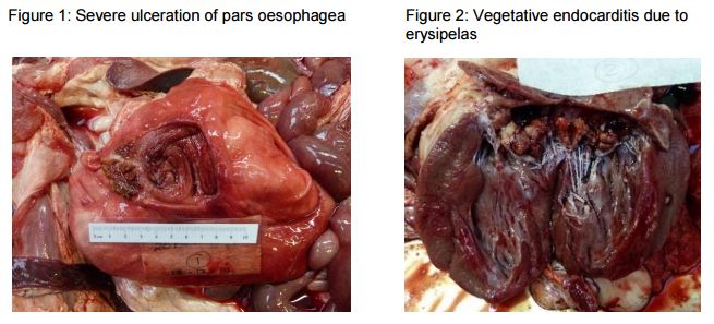
Erysipelas was diagnosed at the University of Bristol in two Gloucester Old Spot gilts, from a group of 13 pigs on a small holding. Both pigs had a short history of malaise and apparent abdominal pain.
At postmortem examination, one gilt had a large haemorrhaging gastric ulcer at the pars oesophagea and the stomach was full of blood (Figure 1).
The other gilt had a smaller non bleeding ulcer, but both had severe chronic vegetative lesions on the left atrio-ventricular valves (Figure 2) from which Erysipelothrix rhusiopathiae was isolated.
Stomach ulceration is not a specific manifestation of erysipelas and in this case it is possible that illness due to endocarditis affected the sows’ appetites and contributed to ulcer formation.
Maintaining vaccination of pigs against erysipelas is an important routine preventative measure and unfortunately the gilts in this small herd had not been vaccinated though the older sows had been.
Iron deficiency anaemia in wasting piglets on a smallholding
Iron deficiency was diagnosed as the cause of ill thrift and lethargy in two piglets submitted to Sutton Bonington from a small open farm with five breeding sows. The holding reported that around a third of piglets in the last two litters had shown poor growth.
One piglet submittted live had very pale mucous membranes and haematology confirmed profound anaemia with a red blood cell count of 1.82 x10¹²/l (reference range 6-9). The carcase showed profound pallor and watery blood.
Biochemistry revealed iron deficiency. Additional laboratory testing did not identify any other concurrent aetiologies for the anaemia and advice was given on provision of iron supplementation.
Nervous Diseases
Severe outbreaks of bowel oedema causing sudden death and nervous signs
Two diagnoses of bowel oedema were made by Sutton Bonington, both quite severe and both associated with Escherichia coli strain E4 (serotype O139:K82) which is recognised as being associated with bowel oedema.
In the first incident, the disease caused 50 deaths from a group of 300 pigs over a three-day period with a clinical presentation of collapse and nervous signs within 24 hours. Disease affected 14-week-old pigs on a 1,000-head finishing unit.
At post-mortem, the eyelids of the submitted pigs were noted to be swollen.
A red fibrin deposit was noted over the small and large intestinal serosa and the distal half of the small intestine contained dark, haemorrhagic contents indicating enteric pathology in addition to nervous disease.
Other organ systems were grossly unremarkable and intestinal bacteriology recovered a heavy growth of haemolytic E.coli strain E4.
In the second incident, bowel oedema was diagnosed following the submission of two dead pigs from a smallholding. The pigs were six-months-old and had been clinically normal the previous day.
On the morning of submission, the owner found three pigs were dead and four others were showing varying degrees of ataxia and nervous signs, including seizures and high pitching squealing.
Both submitted pigs had markedly swollen eyelids and subcutaneous tissues at the extremities were also oedematous.
The intestinal mesentery was oedematous and the content of the distal small intestine was liquid with a hint of haemorrhagic streaking.
A heavy growth of haemolytic Escherichia coli.was recovered from the intestines of both pigs and was serotyped as E4.
Musculoskeletal Diseases
Bacterial joint infections with underlying PRRS
Two 12-week-old pigs were submitted to Thirsk from a nursery-finisher unit to investigate “pigs going off their legs”.
Severe chronic polyarthritis was found in both pigs with some pleuritis, pericarditis and pneumonia.
Trueperella pyogenes was isolated from the lungs and the joints from one pig and Streptococcus dysgalactiae equisimilis from the lungs and joints of the other.
PRRS was detected by PCR on the spleens and, given its immunosuppressive effect, was likely to be a significant factor underlying the bacterial infections diagnosed.