Emerging Threats Quarterly Report – Pig Diseases - July-September 2013
Among the highlights of the latest quarterly surveillance report for the third quarter of 2013 from AHVLA are a Klebsiella septicaemia outbreak in weaned pigs, sporadic haemorrhagic disease, severe polyarthritis due to Strep suis and the detection of an avian-like H1N1 swine flu virus strain in UK pigs. 30 December 2013
 13 minute read
 By:
By: Highlights
- Klebsiella species septicaemia outbreak in post-weaned pigs
- Sporadic haemorrhagic disease in negated swine fever report case
- Severe polyarthritis due to Streptococcus suis type 17
- Avian-like H1N1 swine influenza strain detected again in GB pigs
Ongoing Emerging Disease Investigations
Klebsiella species septicaemia outbreak in postweaned pigs
The second 2013 outbreak of Klebsiella pneumoniae subspecies pneumoniae (Kpp) septicaemia was diagnosed at Bury St Edmunds as the cause of sudden death of 13 30-day-old pigs weaned two days before death. The pigs were found dead on the morning of submission in different tents on an outdoor nursery unit onto which weaners were introduced from two sow herds.
This was the first diagnosis of an outbreak of Kpp septicaemia in post-weaned pigs rather than pre-weaned pigs since the disease emerged in the East Anglian region in the summer of 2011 and the second diagnosis of the disease in 2013.
Neither of the sow herds from which pigs were sourced had diagnosed outbreaks of Kpp septicaemia in pre-weaned pigs. A follow-up submission of pre-weaned pigs dying suddenly on one of the breeding units was found to be due to Glässer’s disease with swine influenza, not Kpp septicaemia. A farm visit was undertaken to obtain background details.
Further information on Kpp septicaemia is available on this link: http://www.defra.gov.uk/ahvla-en/publication/pig-survreports.
All outbreaks of Kpp septicaemia have been diagnosed in the East Anglian region and have consistently presented as sudden deaths of pigs in good body condition during the summer months as illustrated in Figure 1.
Postmortem findings are similar to other causes of septicaemia and laboratory investigation is essential to make a specific diagnosis and distinguish Kpp septicaemia from other septicaemias such as Streptococcus suis or erysipelas.
Cultures for Kpp have been performed on nasal and tonsil swabs collected from pigs submitted to Bury St Edmunds and Thirsk for diagnostic post-mortem examination. Kpp isolates have been obtained from a number of these and, of those analysed so far, no outbreak associated Kpp strains have been identified from pigs which are not from herds with Kpp outbreaks, the last batch of Kpp isolates are being analysed at the end of 2013.
The disease was highlighted at a presentation in May to UK and European pig veterinarians (Bidewell and others, 2013) to maintain awareness of this emerging seasonal disease which appears to be associated with a particular Kpp strain. The disease has been, to date, of low prevalence. So far, the Kpp strain detected in herds with outbreaks has not been detected in pigs from other herds, this work continues.
Sporadic haemorrhagic disease in negated swine fever report case
In August 2013, a case of haemorrhagic disease occurred in an individual adult pig submitted to AHVLA as a diagnostic case. Due to the similarity of the lesions to swine fever, the Veterinary Investigation Officer reported the case as suspect notifiable disease to AHVLA.
The pig was from a small herd of mixed breeds which was put under restriction, the submitted pig and others in the herd were sampled and swine fevers were ruled out by testing. One pig was affected while in-contact pigs were healthy and not pyrexic.
This case superficially resembled those reported in a case series on haemorrhagic disease occurring in single pigs and resembling swine fever (Bidewell and others, 2013). Most of these cases have occurred in non-commercial pigs, one was diagnosed as Border disease (BD) which was ruled out in this pig by testing for both BD and Bovine Virus Diarrhoea viruses by PCR, no other viruses were detected using a virus microarray. Histopathology revealed marked megakaryocytic aplasia explaining the haemorrhagic lesions seen and, with other findings, suggestive of acquired amegakaryocytic thrombocytopenia, an uncommon condition usually associated with a dysfunction of the immune response following an infection.
No involvement of porcine reproductive and respiratory syndrome (PRRS) virus or porcine circovirus-2 (PCV2) was detected. One of the previous cases described by Bidewell and others (2013) in a Saddleback had similar pathology also considered likely to be due to an immune-mediated reaction.
Whilst the absence of clinical signs, lesions and pyrexia in the in-contact pigs provided clinical grounds for negating swine fever, the possibility of a low virulence pestivirus (CSF, BDV, BVDV) meant that swine fever testing was required in this case.
The epidemiological and pathological findings of this case will be added to the case series. The cases have been included in training presentations on porcine notifiable diseases for veterinarians to raise awareness of this clinical presentation and the differential diagnoses to consider.
 |
 |
|
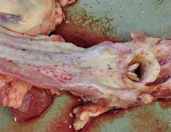
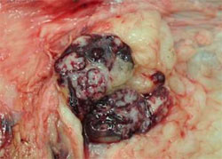
Figure 2 a) Multifocal haemorrhages on mucosa of larynx, pharynx and oesophagus 2 b) lymph node showing mainly medullary dark red-purple discolouration in negated swine fever report case
|
|
Monitoring penicillin sensitivity in Streptococcus suis isolates from pigs
Previous 'Emerging Threats' reports have described the detection of three clinical isolates of Streptococcus suis from pigs with raised penicillin Minimum Inhibitory Concentration (MIC) values above the human clinical break-point for S. pneumoniae central nervous system (CNS) infection. These isolates were from 2009 and 2010, and the most recent one was from March 2013. This has been investigated further by periodic E-test penicillin MIC testing of AHVLA clinical S. suis isolates and a further batch of 38 from 2013 were tested.
The majority had low MIC values <0.05mg/l while two isolates (serotypes 14 and 1/2) had raised MIC values of 0.25mg/l and 0.125mg/l, repectively, but did not exceed the human clinical break-point for central nervous system (CNS) infection (>0.25mg/l). One serotype 1 isolate from June 2013 had an MIC of 0.5mg/l.
There is limited published data available with which to interpret these antimicrobial sensitivity findings. The current medical (BSAC) guidelines do not provide specific values for S. suis but give a clinical break-point in Man of resistant >0.25mg/l for beta-haemolytic streptococci.
The June 2013 isolate is therefore resistant by those criteria and the MIC should be considered as significantly raised at least. It is possible that at this MIC, infection of viscera (for example, lung) with this organism may respond to penicillin, but, because of the blood brain barrier and pharmacokinetic and pharmacodynamic characteristics of the antimicrobial, that CNS infections may not.
In the recent June 2013 case, the S. suis was isolated from the lungs of post-weaned pigs diagnosed with S. suis septicaemia and may therefore have responded to penicillin treatment.
Periodic testing of AHVLA S. suis isolates will continue to determine whether there is emergence of penicillin resistance. The detection of a few isolates with raised MICs highlights the need to maintain this surveillance.
Unusual Diagnoses or Presentations
There were a number of unusual diagnoses this quarter; details of these have been included in monthly AHVLA and SACCVS reports and AHVLA highlights to BPEX, BPA and Pig Veterinary Society. These will be kept under review to assess whether they justify initiation of emerging disease investigations.
Severe polyarthritis due to Streptococcus suis type 17
Nine-week-old pigs were submitted to Thirsk to investigate severe lameness and meningitis on a newly established unit operating within a three-site production system.
Affected pigs were from the first batch to have been weaned and showed severe joint swelling and nervous signs suggestive of meningitis. Sixty pigs from a group of 400 required treatment with injectable penicillin and there was some response to treatment although mortality rate in affected pigs was around 40 per cent.
In submitted pigs there was marked fibrinous arthritis and engorgement of meningeal blood vessels. Streptococcus suis serotype 17 was isolated from joints and brains and severe acute to subacute purulent meningitis was confirmed histologically.
This S. suis type is not commonly associated with primary disease in AHVLA submissions but the profile of pathogens causing disease on newly established "young" herds does sometimes differ from that in established "older" herds.
Bacterial ear infections in outdoor gilts showing head tilt
An unusual outbreak of head tilt, followed by recumbency and mortality or euthanasia, was investigated by submission of a typically-affected 30-week-old gilt which had died to Bury St Edmunds. T
he problem was occurring in a group of purchased gilts introduced onto an outdoor breeding unit. Four of 75 gilts were affected over a three-week period following arrival, and the course of disease in each individual gilt lasted a few days.
Postmortem examination revealed a severe fibrinous pericarditis, and Pasteurella multocida was isolated in septicaemic distribution, including from the meninges, from which Trueperella pyogenes was also isolated. Brain histopathology revealed abscessation involving a linear abscess in the medulla with adjacent purulent encephalitis and subacute to chronic purulent meningitis. The characteristics and location of the purulent encephalitis were consistent with extension from a middle ear abscess.
It was not clear which bacterial pathogen isolated was the primary cause. Streptococcal infections have been associated with outbreaks of middle ear infection, and Streptococcus suis type 1 was isolated from the lung of the gilt but not the meninges.
Mixing, fighting and joint infections are reported predisposing factors for middle ear disease. It may be relevant that another gilt was reported to be lame, and it is possible that fighting occurred following arrival of the gilts on the unit.
Other predisposing factors for middle ear disease can include mange, skin trauma and greasy pig disease, none of which were present in the submitted gilt in which the external auditory canals showed no lesions. There was no evidence of swine influenza or PRRS virus infections.
Necrotic pharyngitis and tonsillitis in growing pigs
A four-month-old pig was submitted to Sutton Bonington from a small breeding and rearing unit to investigate an outbreak of respiratory disease.
Four pigs were affected in three separate groups, there had been no recent changes of note in terms of management.
Marked inflammatory changes were visible in the upper respiratory tract, larynx and pharynx, including the tonsils, with a fibrinous pericarditis. Streptococcus suis type 21 and Haemophilus parasuis were isolated and considered likely to be involved in the pathology. Histopathology on a range of tissues did not suggest viral involvement.
In a second case, 11-week-old pigs were submitted to Bury St Edmunds to investigate ill-thrift in 10 of a batch of 500 kept in large groups on deep straw.
Gross lesions in one of the pigs were typical of salmonellosis with necrosis of the distal small intestine and large intestine and yellow diarrhoea and this diagnosis was confirmed. In the second pig, gross lesions were unusual with a ring of blackened necrosis involving the proximal oesophagus, pharynx and part of the tonsils. Fusobacterium necrophorum was isolated together with Streptococcus suis type 16 from the necrotic areas. The anaerobe was likely to be most significant with respect to the lesions (this organism is also the cause of calf diphtheria). Histopathology supported a bacterial cause for the lesions.
The continuous nature of this nursery-finisher unit, together with the frequent entry of large batches of pigs kept in big groups with minimal opportunity for effective cleaning and disinfection between batches were likely predisposing factors leading to salmonellosis on the unit. The Fusobacterium species infection was also considered likely to reflect poor hygiene.
Previous cases of necrotic tonsillitis have been seen in pigs with severe porcine circovirus 2-associated disease (PCVAD) concurrent with PRRS, however, testing for PRRS and histopathology for PCVAD did not reveal involvement of these pathogens in either of these cases. The detection of necrotic tonsillitis can be of concern as the lesion may be seen in Aujeszky’s disease – disseminated multifocal necrosis is a feature of herpes virus infections.
Changes in Disease Patterns and Risk Factors
Avian-like H1N1 swine influenza strain detected again in GB pigs
Swine influenza virus was detected in seven outbreaks between July and September 2013 using the m gene PCR in spite of the reduced submission numbers.
Pandemic H1N1 2009 (pH1N109) and H1N2 strains remained predominant in this quarter but avian-like H1N1 was detected in August in one herd in the Thirsk region. This strain was last identified in November 2011, also in the Thirsk region.
The emergence of pH1N109 in pigs from 2009 is considered likely to be involved in the decline in avian-like H1N1 detection as pH1N1 has replaced it as the predominant H1N1 strain. The m gene PCR is more sensitive than virus isolation and it is not always possible to identify the virus strain in samples where the Ct value is high in the PCR.
The strain involved in outbreaks of swine influenza was successfully identified in five of the seven m gene-positive submissions. This is an improved proportion compared to the last quarter reported in the Q2 2013 'Emerging Threats' report. Tissue culture is being used in addition to conventional virus isolation in eggs in selected cases.
Influenza in pigs, and its contribution to porcine respiratory disease complex and to the evolution of novel strains remains a concern. and the availability of the Defra-funded swine influenza surveillance (www.defra.gov.uk/ahvla-en/files/pub-vet-si.pdf) remains an important means of surveillance.
Tiamulin-resistant Brachyspira hyodysenteriae isolate
Another tiamulin-resistant isolate was identified during this quarter through the routine tiamulin MIC testing funded from the 'Monitoring of Antimicrobial Resistance in Bacteria from Animals and their Environment Project' within AHVLA.
The isolate was from a September submission to Thirsk from a small outdoor breeding herd. The MIC value for the B. hyodysenteriae isolate was 16mg/l tiamulin hydrogenfumarate, which is above the MIC break-point for tiamulin against B. hyodysenteriae generally considered to be >4ug/ml for agar dilution.
The development of resistance of Brachyspira hyodysenteriae to antimicrobials commonly used in the control of swine dysentery is a recognised risk, particularly in situations where medication is used long-term.
Control of swine dysentery using alternative interventions (all-in, all-out management systems; cleaning and disinfection; and partial and total depopulation leading to eradication) is vital to prevent the development of wider antimicrobial resistance but may not be effectively undertaken in small non-commercial units.
The isolate will be tested free of charge against other antimicrobials licensed for treatment of swine dysentery as part of AHVLA surveillance for multidrug resistant B. hyodysenteriae.
Horizon-scanning
Neonatal Porcine Diarrhoea Syndrome
Since 2008, a problem known as Neonatal Porcine Diarrhoea syndrome (NNPDS) was reported in certain European countries, including Denmark and France, of diarrhoea in neonatal pigs differing from usual outbreaks in being unresponsive to antibiotics and common management practices. Routine laboratory examinations have not detected any pathogen related to this syndrome.
Currently, most Danish pig veterinarians see cases in a proportion of the herds they attend. A systematic study was undertaken on four well-managed Danish herds suspected to suffer from New Neonatal Porcine Diarrhoea Syndrome (Kongsted and others, 2013).
The herds were selected according to certain criteria:
- the presence of diarrhoea responding poorly to antibiotics during the first week of life (at least 30 per cent affected litters for a period of minimum 6 months)
- routine vaccination of sows against E. coli and Clostridium perfringens type C
- failure of preventive management interventions
- negative results in routine diagnostic examinations for enterotoxigenic E.coli, Clostridium perfringens type C and rotavirus in five diarrhoeic piglets aged one to four days; and
- PRRS-negative by testing.
Based on the findings, the following case-definition of NNPDS was suggested: “non-haemorrhagic diarrhoea during the first week of life, without detection of known infectious pathogens, characterized by milk-filled stomachs and flaccid intestines at necropsy".
The predominant histological lesions were villous atrophy in jejunum and ileum. Examination for coronavirus was by use of pan-corona RT-PCR assay and none was detected, suggesting that porcine epidemic diarrhoea virus (PEDv) is not involved in NNPD.
The DNR for enteric syndrome in neonatal pigs to Q3, 2013 was 10 per cent compared to 27 per cent in prior years, providing no evidence that NNPDS is emerging as a pig health issue in GB pigs at present.
Further Reading
You can view the full report by clicking here.
Find out more about the diseases mentioned by clicking here.
December 2013