


AHVLA Reports First Swine Dysentery Cases for Two Years in May 2014
UK - Among the key findings in the AHVLA Pig Disease Surveillance Monthly Report for May 2014 are fungal and bacterial placental infections result in abortions; the first swine dysentery outbreak diagnosed in East Anglian region since 2012; enteric disease and deaths due to PCV-2 and oedema disease outbreak in weaners with meningitis-like signs.Reproductive Disease
Fungal cause of abortions and weak neonates
Fungal disease was diagnosed when weak neonatal and aborted piglets were submitted to Bury St
Edmunds from two of four sows affected on a 1,000 sow outdoor breeding unit. The four sows were from different parities and were well and in good body condition. This was the first batch affected in this way, although the last few batches of sows to farrow had significantly reduced litter sizes. There were no significant foetal lesions visible grossly but the two placentas submitted appeared thickened and wrinkled with focal haemorrhage, and as shown in Figure 1. Aspergillus fumigatus was isolated from several foetal stomach contents and from the placentas and histopathology supported these culture findings with lesions of a mycotic placentitis and a purulent bronchopneumonia in one piglet as a likely sequel to the fungal infection. An environmental source of infection was suspected with bedding or stacked straw being possible sources.
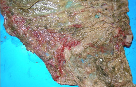
Bacterial placentitis causes abortion
Unusual diagnosis of bacterial placentitis due to Bacillus species was suspected by Bury St Edmunds to be the cause of abortion on an outdoor breeding unit. Two sows had aborted from a batch of 90 13-weeks in-pig. The aborted foetus was from a gilt and was the only foetus found although three placentas were present indicating a larger litter had been aborted. The problem of around two abortions per farrowing batch has occurred in the last three batches and around the same time. Normal numbers of live piglets were being delivered with a slight increase in stillbirths, and mummies were rare. Piglet viability was good although there has been long-term variation in piglet size within litters, possibly due to larger litter sizes. No other problems were reported on the unit or rearing farms it supplied. There was gross evidence of reddening and thickening in the placentas not dissimilar to that seen in the case of fungal placentitis described above but profuse growths of both Streptococcus porcinus and Bacillus species were obtained from three placentas with no fungal growth. Histopathology confirmed a bacterial placentitis and the morphology of the organisms associated with the lesions suggested that the Bacillus species was most likely to be the significant organism. No other infectious agents of abortion were detected.
Alimentary Disease
First swine dysentery outbreak diagnosed in East Anglian region since 2012
Swine dysentery was confirmed on a breeder-finisher unit when two live 12-week-old pigs were submitted to Bury St Edmunds to investigate diarrhoea in approximately 50 per cent of 10 to 12-week-old pigs in strawed pens with no deaths. Mucus and, in just two faeces, blood flecks had been noted in the diarrhoea on farm. Diarrhoea was first noticed in finishing pigs and was reported to have been on-going for approximately six to eight weeks. A scrape-through system is operated on the unit. The two untreated pigs were submitted live with grey-green diarrhoea. In one, the large intestine was thickened and the mucosa reddened along its entire length with accumulations of mucus adherent to the mucosa (Figure 2), raising a suspicion of colitis due to Brachyspira species. The colon of the second pig showed thickening of the colon with a pale mucosa and no mucus visible. Brachyspira hyodsenteriae was isolated from both pigs and was detected by PCR.
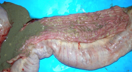
The Brachyspira hyodysenteriae isolate was found to be sensitive to tiamulin. AHVLA made no diagnoses of swine dysentery in East Anglia in 2013 and only one, in smallholder pigs, in 2012.
More information is available on swine dysentery from BPEX on this link: www.bpex.org.uk/2TS/health/swinedysentery.aspx.
Wasting due to post-weaning E.coli infection
Enteric colibacillosis was diagnosed at Bury St Edmunds as the cause of wasting from approximately 10 days postweaning occurring over a six-month period on a 240-sow indoor commercial breeder-finisher weaning weekly into slatted rooms. Up to 20 per cent of pigs were estimated to be affected with around a quarter of these being culled or dying. Pigs were vaccinated for Mycoplasma hyopneumoniae and PCV2 at weaning. The rest of the herd is performing well with good conception and farrowing rates. Three live five and a half week-old pigs were submitted which had not been treated. The pigs were alert but in poor body condition and had diarrhoea. The large intestines in particular were dilated, thin-walled and filled with excessive watery light-green contents as illustrated in Figure 3 and E.coli strain V17 (serotype O157:KV17) was isolated in heavy pure growth. Rotavirus was also detected in one of the pigs and is not uncommonly detected concurrently with enteric colibacillosis immediately post-weaning. Porcine epidemic diarrhoea virus was not detected.
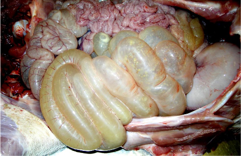
Neonatal piglets with watery diarrhoea due to likely clostridial enterotoxaemia
Clostridial enterotoxaemia was strongly suspected from detection of alpha toxin and histopathology to be the cause of watery diarrhoea affecting neonatal piglets in a third of litters in a single parity herd at its fourth parity. In affected litters, approximately one third of piglets showed signs and there was a mixed response to antimicrobial treatment with some piglets recovering. Three dead piglets were submitted with gastric necrosis and yellow watery diarrhoea. Gammaglobulin estimation revealed less than adequate colostral antibody uptake which will have predisposed to disease if piglets were exposed to a contaminated environment.
Wasting and diarrhoea due to both ileitis and colitis in finishers
Concurrent porcine intestinal adenomatosis and Brachyspira pilosicoli colitis were diagnosed at Bury St Edmunds as the cause of diarrhoea and wasting over a three-week period in one batch of 16-week-old pigs. Twenty of 170 were reported to be affected with four deaths. The pigs were kept in strawed pens and managed on a continuous basis. No vaccinations are used in the rearing pigs on this breeder-finisher PRRSv-negative unit and there was concern that porcine circovirus 2-associated disease (PCVAD) was involved which had never been diagnosed on the unit. Three euthanased untreated pigs were submitted in which the most significant gross pathology involved the small and large intestines with thickening, corrugation and necrosis (Figure 4).
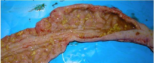
Involvement of porcine intestinal adenomatosis/necrotic enteritis due to Lawsonia intracellularis was supported by the detection of MZN-stained curved organisms in smears and confirmed by histopathology which also ruled out PCVAD. Brachyspira pilosicoli was also isolated and histopathology indicated its likely involvement in the large intestinal lesions in at least one pig. As a secondary, but still significant finding, in all three pigs there was cranioventral consolidation of variable severity. No viruses were detected and histopathology revealed chronic bronchointerstitial pneumonias consistent with mycoplasmosis with Mycoplasma hyopneumoniae detected. The unit is known to be Mycoplasma hyopneumoniae positive but, unusually, does not vaccinate growing pigs.
Enteric colibacillosis with rotavirus in neonatal piglets with watery diarrhoea
Neonatal piglets less than two-days-old were submitted to Thirsk to investigate diarrhoea starting within one day of birth. Post-mortem examination confirmed an enteritis with intestinal contents being yellow and watery and with slight reddening of the intestines. Cultures revealed the presence of E. coli serotype 0139:K82 (E4) and rotavirus was also detected in one of the piglets. The isolated E. coli is a known enteropathogenic strain associated with enteric disease and also oedema disease in older pigs. Porcine epidemic diarrhoea virus was not detected.
Respiratory Disease
PRRS virus field challenge detected in finishers
Porcine reproductive and respiratory syndrome virus (PRRSv) was detected by PCR in sera from 15-week-old pigs of which around 10 per cent were showing increased coughing on an indoor breeder-finisher. This detection indicates field challenge with PRRSv but further testing, by post-mortem examination, would be required for complete diagnosis. No antibody was detected to swine influenza virus.
Respiratory disease and deaths due to Actinobacillus pleuropneumoniae
Respiratory signs affecting over half of 160 14-week-old finishers three weeks after entering a multi-source indoor finishing unit were investigated by submission of two pigs found dead. Both had dark red cranioventral consolidation with fibrinous pleurisy and Actinobacillus pleuropneumoniae was isolated from both, confirming it as the cause of death. No underlying viral involvement was detected in the submitted pigs. Only one of the three sources on farm was affected in one shed at the time of submission and it is possible that differing pathogen and immune status in the three sources led to disease in one.
Systemic Disease
Enteric disease and deaths due to porcine circovirus-2 associated disease
Two pigs aged 10 and 13-weeks were submitted to Shrewsbury to investigate the cause of diarrhoea and increased sudden deaths on a 300-sow breeder-finisher unit. The farm had previously vaccinated weaners for PCV2 but stopped about two years ago. Six pigs had died within a three-week period. The younger submitted pig was markedly diarrhoeic and had minor lung consolidation. Histopathology and immunohistochemistry confirmed porcine circovirus-2 (PCV-2) associated disease (PCVAD) and this was considered the primary cause of disease and death in this pig. The older pig had enlarged, pale, firm kidneys with fine petechiation over the kidney cortices. Histopathology confirmed a severe glomerulo-nephritis, with pathology typical of porcine dermatitis and nephropathy syndrome (PDNS), likely to be associated with the PCVAD occurring on farm. Since obtaining the diagnosis, the farm has started vaccinating weaners for PCV2 again.
Erysipelas suspected in replacement breeding gilts
Erysipelas was strongly suspected by Shrewsbury when a gilt was submitted to investigate illness in two in a group of eight gilts approximately six months of age on a unit of 40 gilts and sows. The gilts had been only recently completed their primary course of vaccination for erysipelas and a few days later two developed purple ecchymotic haemorrhages on the ears and the hind quarters and became pale, lethargic and inappetant. There was some improvement following antimicrobial treatment, but one died and was submitted. A proliferative valvular endocarditis was found, consisting of large nodules on the left atrioventricular valve. Focal inflammatory lesions in the kidneys and lungs were histopathologically consistent with secondary embolic lesions. No bacteria were isolated, probably due to prior antibiotic treatment; such lesions in pigs are usually associated with either Erysipelothrix rhusiopathiae or Streptococcus suis infection.
Nervous Disease
Streptococcal meningitis and septicaemia in growers
Ten deaths were reported in nine -week-old pigs on a unit with over 2,500 growing pigs, with others showing neurological signs and responding to antimicrobial treatment. Two pigs submitted had fibrinous peritonitis and pericardial effusions, and one also had pneumonia and a purulent meningitis. Streptococcus suis type 2 was isolated from the brains of both pigs and the liver of one, consistent with a diagnosis of streptococcal meningitis and septicaemia.
Oedema disease outbreak in weaners with meningitis-like signs
Six-week-old pigs were submitted to Thirsk to investigate nervous signs and mortality over a weekend not responding to treatment for suspected meningitis. Affected pigs were reported to have sudden onset eyelid swelling and a proportion then developed meningitis like signs. The indoor unit operates a single source batch system with pigs entering every five weeks. Two of the pigs were in lateral recumbency and paddling with nystagmus and slightly swollen eyelids. Post-mortem examination revealed subtle subcutaneous oedema, a wet appearance to subcutaneous tissues and oedema of the mesocolon in two of the pigs. None of the pigs were pyrexic which, together with the clinical history and gross findings, supported a suspicion of oedema disease due to E. coli rather than a bacterial meningitis. Haemolytic E. coli (serotype 0139:K82, strain E4) was isolated from small intestinal contents from an untreated pig with marked signs. This strain can be associated with both enteric disease and oedema disease in pigs.
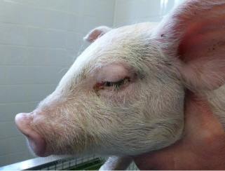
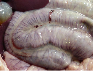
Histopathological examination of two brains revealed oedema and an associated encephalopathy supporting the diagnosis of oedema disease. PRRS genotype 1 (European strain) was also detected in pooled sera from these pigs.
There has been an increasing trend in oedema disease as a cause of porcine nervous disease in AHVLA submissions over the last 12 months and it is worth bearing in mind the possibility of oedema disease even when oedema of the subcutis or organs is absent. Bacterial meningitis (mainly due to Streptococcus suis, some due to Haemophilus parasuis) remains the predominant AHVLA diagnosis in porcine nervous disease in the 12 months to the end of March 2014, with water deprivation, oedema disease, middle ear disease, brain abscessation and porcine sapelovirus infection making up most of the remainder of the diagnoses.


