AHVLA Reports Neonatal Pig Deaths from Klebsiella in July 2014
UK - The Animal Health Veterinary Laboratories Agency (AHVLA) has reported neonatal deaths due to clostridial enterotoxaemia, the first outbreak of Klebsiella pneumoniae septicaemia outside East Anglia, polyuria, polydipsia and inappetance in adult sows due to vitamin D toxicity in its July 2014 Surveillance report. 13 October 2014
 8 minute read
 By:
By: Alimentary Disease
Clostridial enterotoxaemia causing neonatal diarrhoea and deaths on outdoor units
Two incidents of neonatal diarrhoea due to clostridial enterotoxaemia were diagnosed at Bury St Edmunds on unrelated outdoor breeding units. Both involving gilt litters. In the first, one-day-old live piglets were submitted to investigate diarrhoea affecting neonatal piglets in gilt litters since just after Christmas.
In the first case, 10 of 35 gilt litters were affected with neonatal mortality increasing in these litters from around 12 to 20 per cent. Neonatal mortality in the rest of the herd was just seven to eight per cent. Gilts were homebred with the gilt paddocks sited approximately one mile from the dry sow area. Congenital tremor-type A2 was diagnosed in March 2014 in gilt litters and a few cases were still being seen. In two piglets submitted there was a very scant quantity of yellow, slightly frothy fluid in the small intestines.
The mucosa of the duodenum and proximal jejunum was white in colour and moderately thickened with a fluffy appearance. The caecum and colon contained a scant yellow fluid and the colonic mesentery was oedematous. Testing for neonatal enteropathogens detected alpha clostridial toxin in small intestinal contents of one piglet and gammaglobulin estimation was suggestive of less than optimal colostral antibody transfer in both. Intestinal histopathology supported the detection of alpha toxin and was consistent with clostridial enterotoxaemia. In the second incident, which had been ongoing for three to four months, diarrhoea was occurring from one-day-old followed by wasting in piglets which survived.
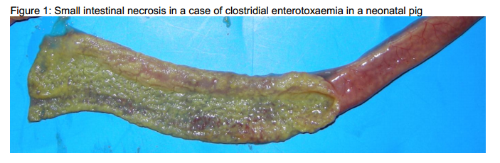
Ten litters of each batch of 70 gilts farrowing weekly on the single parity unit were affected. Not all piglets in a litter were affected, but a poor response to antimicrobial treatment was reported, with an estimated 50 per cent case mortality. A one-day-old piglet submitted had jejunal thickening and necrosis (Figure 1) suggestive of clostridial enterotoxaemia.Alpha toxin was detected in small intestinal contents from this piglet.
A four-day-old piglet submitted was in very poor body condition, severely dehydrated with urate accumulations in the kidneys and was considered to be a more chronic case. Gammaglobulin testing in the one-day-old piglet revealed hypogammaglobulinaemia and this, together with possibly inadequate
Respiratory Disease
Widespread respiratory disease of mixed bacterial and viral aetiology
Three pigs were submitted to investigate widespread sneezing, respiratory disease, a few cases of meningitis, five per cent mortality and some wasting in 1,300 seven-week-old pigs from a single source on an indoor nursery-finisher unit.
The pigs were vaccinated on arrival for Mycoplasma hyopneumoniae and PCV2. Pigs had been treated for meningitis and diarrhoea since arrival and there was unevenness in the batch. All three pigs submitted were in poor body condition. Two had a fibrinous polyserositis and Pasteurella multocida was isolated from the lungs of one of these pigs. Cultures for Haemophilus parasuis did not yield the organism but antimicrobial treatment may have affected culture results and its involvement cannot be ruled out. No evidence of PCV2-associated disease (PCVAD) was found in these two pigs. However, the third pig had pulmonary oedema, bronchointerstitial pneumonia and a marked pleural effusion suggestive of the acute pulmonary oedema presentation of PCVAD which was confirmed by histopathology and immunohistochemistry. Although PCVAD was not the predominant diagnosis in this submission, the PCV2 was genotyped and was typical of PCV2b strains found previously in GB pigs. No swine influenza or PRRS involvement was found.
Systemic and Miscellaneous
First outbreak of Klebsiella pneumoniae septicaemia outside East Anglia
Two submissions of 10- to 12-day-old piglets from the same farm to Starcross resulted in the first diagnosis of Klebsiella pneumoniae subsp. pneumoniae (Kpp) septicaemia outside of the East Anglian region. The post-mortem findings were very similar to those seen in previous cases with piglets in good condition showing evidence of recent feeding but with a septicaemic appearance and marked haemorrhages on the intestinal serosa.
The Kpp organism was isolated from a wide range of internal sites. The outbreak was typical of most previous ones, with unexpected deaths of preweaned pigs in good body condition on an outdoor breeding unit. However, mortality was significantly higher at 16 per cent over a period of four weeks. Antimicrobial treatment was instituted at the same time as iron supplementation and, when treated pigs came through, mortality stopped. This treatment will be continued until the autumn as, so far, Kpp outbreaks have been confined to the summer.
The herd has been closed for several years and litters from sows of all parities were affected, the reason for the outbreak is not known but the isolate will be typed to determine whether it is the same as the emerging ST25 strain detected in previous Kpp outbreaks. As the presenting sign of sudden death is not specific for Kpp septicaemia, diagnosis depends on post-mortem examination and bacterial culture.
Porcine circovirus 2-associated wasting in unvaccinated pigs
A dead 10-week-old piglet was examined at Penrith. It was the second reported loss from a group of 20 pigs with a history of malaise and loss of body condition. The pigs had been purchased as weaners at four-weeks-old and were kept outdoors, they were not known to have been vaccinated for PCV2. A severe pneumonia, lymphadenopathy and hepatitis were found and histopathology and immunohistochemistry confirmed PCVAD with widespread staining of PCV2 antigen in intestines and lymph nodes. This emphasises the continued value of PCV2 vaccination.
Two different presentations of erysipelas in unvaccinated pigs
Two diagnoses of erysipelas were made in July by Bury St Edmunds on unrelated units. One was an outbreak of septicaemia in three to four-week-old pre-weaned pigs on a small unit with 43 pigs. Four deaths had occurred from one litter with piglets becoming listless prior to death. One dead piglet was submitted in which there were non-specific lesions with skin reddening, fibrin stranding and slight yellowing of the subcutis and pure growths of Erysipelothrix rhusiopathiae were isolated from multiple internal sites, confirming the diagnosis.
Erysipelas had been suspected to have also been affecting the sow and vaccination was recommended. The second case diagnosed was in a young breeding boar which had not yet been vaccinated after arriving on farm two weeks earlier.
The boar was found dead with no prior signs. Postmortem examination revealed marked intraperitoneal haemorrhage from a ruptured liver, and congested oedematous lungs reflecting heart failure and passive congestion due to a 3 severe vegetative endocarditis affecting the left atrioventricular valve. Erysipelothrix rhusiopathiae was isolated from the heart valve.
Typical case of streptococcal disease
Approximately 40 per cent of 1,600 eight-week-old growers were reported to be affected with respiratory disease and nervous signs over a two-week period with a poor response to antimicrobial treatment. The piglets were vaccinated against Mycoplasma hyopneumoniae and PCV2 on arrival at the farm. The piglets were housed in groups of 40 per pen. Two dead pigs were submitted, both had pneumonias and one also had fibrinous polyserositis. Streptococcus suis type 2 was isolated from meninges and lungs confirming a diagnosis of streptococcal disease and no virus involvement was detected.
Skin Disease
Outbreak of greasy pig disease in neonatal piglets
Greasy pig disease (exudative epidermitis) was diagnosed at Bury St Edmunds as the cause of skin lesions in several one-week-old litters of one batch of farrowing sows. Piglets developed skin scabs and redness over a few days. Some died, others lost weight and a poor response to antimicrobial treatment was reported.
There was no previous history of greasy pig disease on the unit and the herd was closed with no changes to management of the fully slatted farrowing houses to explain the outbreak of disease. No mange mites were detected. All three piglets submitted had raised multifocal waxy brown scabs and a generalised coating of brown exudate over reddened thickened skin from which Staphylococcus hyicus was isolated in pure growth, confirming the diagnosis which histopathology also supported. The staphylococcal isolate was fully sensitive to antimicrobials tested and early treatment with supportive care is important to limit piglet deaths from generalised disease due to dehydration and toxin absorption.
Polyuria, polydipsia and inappetance in adult sows due to vitamin D toxicity
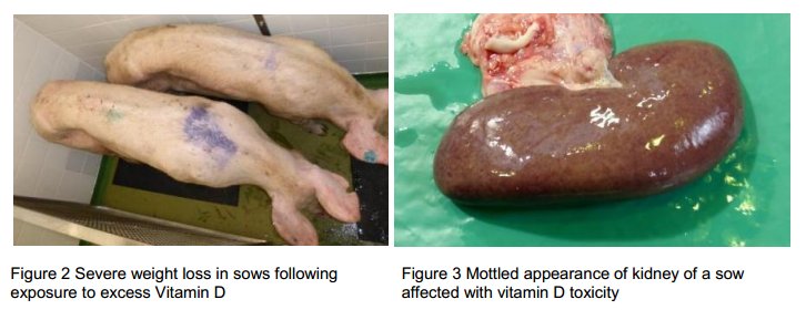
All the sows on two breeding units were affected by vitamin D toxicity due to accidental addition of the vitamin to the mineral pre-mix resulting in marked overdose of vitamin D to the pigs consuming the affected ration. Fortunately, the affected feed was only fed to two pig units and was replaced as soon as concerns were raised with voluntary restriction of cull sows on-farm agreed for a specified period to protect the food chain. Clinical signs included marked polydipsia and secondary polyuria followed by inappetence and weight loss (figure 2). Post-mortem examination of culled sows at Thirsk were principally of a nephropathy as shown in figure 3 which was confirmed by histopathology which revealed calcium phosphate mineralisation. Further details of the case are described on this link:https://www.gov.uk/government/uploads/system/uploads/attachment_data/file/352185/pub-survrep-p0214.pdf
Mortality involving rectal prolapses in finishers
Two 12 to 14-week-old pigs were submitted to Thirsk to investigate ongoing mortality in well-grown pigs. Some pigs were developing rectal prolapses, possibly as a sequel to coughing. The rectal prolapses were being traumatised by pen mates, leading to complications such as peritonitis.
One of the submitted pigs had died from a torsion of the small intestine. The other pig had a rectal prolapse and fatal peritonitis as the distal rectum was detached from the anus and faeces was leaking into the abdominal cavity.
There was clear evidence of trauma to the rectum and anus and this pig also had a pneumonia. Mixed growths of non-haemolytic E.coli and Streptococcus dysgalactiae equisimilis were isolated from the lungs and no viral involvement was detected which may be secondary to bacteraemic spread from the damaged rectal prolapse. Pigs appear to be particularly prone to rectal prolapse and anything which increases abdominal pressure or causes straining can predispose to the condition, for example, diarrhoea, constipation, coughing, low-fibre diets, pigs piling on top of each other due to chilling.
Predisposing factors need to be considered and any likely to be playing a role should be addressed promptly where numbers of pigs are being seen with rectal prolapse.
Possible factors and advice on how to deal with affected pigs from NADIS.
Advice on dealing with casualty pigs is provided in the document from the Pig Veterinary Society.