

AHVLA Report: Piglet Deformities, PRRS, Septicaemia, Endocarditis
UK - The Animal Health Veterinary Laboratories Agency (AHVLA) has reported piglet deformities, unusual Brachyspira species isolated from pigs with mucoid diarrhoea, PRRS, haemorrhage associated with copper deficiency in pigs on milk-based diet and septicaemia and endocarditis in its Pig Disease Surveillance Monthly Report for September 2014.The diagnoses of enteric disease in neonatal and preweaned pigs in the 12 months to the end of September 2014 are shown in figure 1. Rotavirus, clostridial disease and E. coli are relatively equally represented and mainly affect neonatal pigs in the first week of life. Coccidiosis was not often diagnosed. This may, in part, be due to the fact that when samples are submitted rather than pigs, coccidial oocysts may not be detected in the faeces in the first few days of disease. Coccidiosis due to Isospora suis can occur from five-days-old and is mainly seen from seven to 20 days of age. The ideal material for diagnosis of coccidiosis is live untreated affected piglets for post-mortem examination including rapid fixation of the intestines, allowing intestinal histopathology and detection of Isospora suis. No porcine epidemic diarrhoea virus was detected.
Reproductive Disease
Piglet deformities over a short period on an outdoor breeding unit
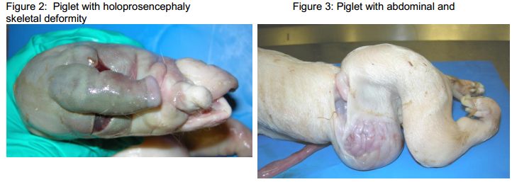
The occurrence of 40 deformed piglets across about 25 litters in a batch of farrowing sows on an outdoor unit was investigated when two were submitted to Bury St Edmunds. One to two piglets were deformed in each of the affected litters which were from any parity of sow. A few deformed piglets had also been seen in each of the previous two farrowing batches. The two submitted piglets had different deformities as illustrated in Figures 2 and 3.
The findings in the piglet with cyclopia and a proboscis represent a severe form of holoprosencephaly (malformations of midline of prosencephalon and related facial/ocular anomalies) which is a recognised malformation syndrome in mammals and has been recorded previously in pigs as far back as 1908. It is usually a sporadic event in pigs. The malformation in this piglet will have occurred prior to day 16 of gestation. The timing is not suggestive of involvement of an infectious agent.
Teratogenic effects are recorded in the foetuses of ewes that have grazed on Veratrum californicum (California corn lily, white or California false hellebore) in North America, and exposure to certain teratogenic drugs and inherited genetic abnormalities can also be involved in holoprosencephaly.
Histopathology on the piglet with abdominal and musculoskeletal deformity again does not suggest involvement of an infectious agent and further examinations are in progress to evaluate the exact timing of the insult. Investigations into this transient event continue. The unit reported that only two litters had deformed piglets in the subsequent batch of sows to farrow. If you hear of other farms experiencing similar problems, AHVLA is interested to hear about these and discuss their submission for investigation. It is also useful if images of the range of deformities seen can be provided.
Alimentary Disease
Further incidents of neonatal diarrhoea due to clostridial enterotoxaemia
Further to similar incidents reported recently, another outbreak of clostridial enterotoxaemia was diagnosed in neonatal piglets which were affected between one and seven-days-old with yellow watery diarrhoea. The problem had been going on for several weeks when mortality spiked to 20 per cent, prompting submission of live affected one-day-old piglets to Bury St Edmunds for investigation. The piglets were diarrhoeic but quite bright and post mortem examination revealed watery small intestinal contents. Clostridium perfringens alpha toxin was detected in one piglet and histopathological lesions in the intestines supported type A enterotoxaemia.
Intestinal torsions in outdoor replacement gilts
Two young replacement gilts which died suddenly were submitted to Bury St Edmunds from an outdoor unit on which four deaths had occurred in the group of 67 soon to be served. One had died of an intestinal torsion, the other from peritonitis and, in both, sandy sediment and stones weighing 10kg were present in the intestines and are likely to have predisposed to the clinical problem in both.
Excessive ingestion of soil, stones and sand can occur in pigs recently introduced to outdoor units and occasionally results in small “outbreaks” of intestinal torsion. Interventions to try and prevent this include more frequent feeding (which was already in place on this unit) and provision of bales of straw or similar to distract the pigs.
Unusual Brachyspira species isolated from pigs with mucoid diarrhoea
Brachyspira hyodysenteriae was isolated from two faecal samples from 14-week-old pigs with mucoid diarrhoea and wasting in the Thirsk region. These faeces gave anomalous results in the Brachyspira PCR and further investigation is in progress at APHA and SRUC to determine the identity of the organism and whether the Brachyspira species isolated are similar to atypical B. hyodysenteriae strains that were detected in the late 1990s which have not been seen in recent years. In the light of these unusual results, further faecal samples from untreated pigs on the affected farm were requested and this organism has not been detected again.
Outbreaks of salmonellosis continue to cause diarrhoea, wasting and mortality
Salmonellosis was considered the likely cause of loss of condition and diarrhoea, together with lesions of diphtheritic colitis in 12-week-old pigs. Monophasic Salmonella Typhimurium-like variant 4,12:i:-, phage type 193 was isolated from a faecal sample submitted – monophasic S. Typhmurium-like variants have emerged to become predominant in pigs in recent years. In a second incident in younger pigs, salmonellosis due to S. Typhimurium U288 was diagnosed in five to six-week-old pigs from an indoor unit on which 30 deaths had occurred since weaning with diarrhoea and wasting affecting around 30 per cent of pigs. Rotavirus was also detected in one of the pigs and was likely to have been of secondary clinical significance. There was intestinal thickening in two of the pigs, a necrotic enterocolitis in one and Salmonella was isolated by direct culture from all three pigs. No enteropathogenic E. coli were isolated. Previous batches of pigs from the same source were reported to have been affected with salmonellosis.
Parasitic disease causing weight loss in a smallholder herd
Oesophagostomum species worm infestation was considered the likely cause of chronic weight loss and eventual euthanasia of an 11-month-old sow which was submitted to Camarthen. She was one of eight sows kept in individual arcs on three acres of land. The sow was found to be thin with profuse numbers of worms in the colon and caecum, identified as Oesophagostomum species. A high faecal worm egg count was detected of 4,400 trichostrongyle-type eggs per gram. In large numbers as found here, Oesophagostomum species can cause condition loss, and infection can build up on land where pigs have been kept for a prolonged period without appropriate control measures including anthelmintic treatment.
Respiratory Disease
Active swine influenza with bacterial diseases in finishers with mixed clinical signs
Active swine influenza infection, Streptococcus suis and Pasteurella multocida were isolated from lung and Salmonella Typhimurium phage type U308 from intestine when samples were submitted by a practitioner from on-farm post-mortem examination. These findings explained the respiratory signs, diarrhoea and weight loss in approximately 10 per cent of 110 fourteen-week-old pigs at a finisher unit on which eight pigs had died. The sampled pig had bronchopneumonia with pulmonary abscessation and was diarrhoeic. Fixed heart was submitted from a second pig and a mild non-suppurative myocarditis was identified, no other samples were available from this pig to investigate further. Virus isolation to identify the strain of influenza virus was not successful but PCR testing showed that it was not pandemic H1N1 2009.
Porcine reproductive and respiratory syndrome (PRRS) outbreaks diagnosed
Porcine reproductive and respiratory syndrome (PRRS) was diagnosed with bacterial lung infections in several units, the immunosuppressive nature of the PRRS virus likely to have played a significant role in the clinical disease.
In one of these outbreaks, respiratory disease due to PRRS with Pasteurella multocida and Glässer’s disease, was diagnosed in housed nine-week-old pigs with respiratory signs, wasting and diarrhoea affecting five per cent of 1500 pigs with two-thirds of those affected dying. The pigs submitted had severe pneumonias and one had a fibrinous pericarditis and pleurisy typical of Glässers which was confirmed by isolation of H. parasuis. One of the pigs also had necrotic large intestinal lesions typical of salmonellosis which was confirmed with the isolation of S. Typhimurium phage type U302. The pigs had been vaccinated at weaning for PRRS, therefore when PRRSv was detected by PCR, immunohistochemistry was performed and confirmed involvement of PRRS in the pneumonia and clinical disease.
PRRS with P. multocida infection was also diagnosed in finisher pigs which were not vaccinated for PRRS when individual lungs were submitted from the abattoir to investigate respiratory disease earlier in rear. There was pale pink cranioventral consolidation suggesting chronic lesions involving approximately 20 per cent of lung fields. PRRS virus was detected in all six lungs submitted and histopathology was consistent with the dual infection detected.
A long-term problem of wasting with some diarrhoea in growers on an indoor breeder-finisher was investigated by submission of three typically affected untreated live seven-week-old pigs to Bury St Edmunds. A previous submission of five-week-old pigs had diagnosed enteric colibacillosis and interventions were put in place to address this. Subsequently, wasting started to be seen in older pigs. In the batch from which pigs were submitted, 25 per cent of pigs were affected with inappetance, wasting and poor response to injectable antimicrobial treatment. Five pigs had been culled or died. Pigs were vaccinated for Mycoplasma hyopneumoniae and PCV2. The submitted pigs were in poor body condition with wheezy respiration and one had a slight left head tilt. All three pigs had severe bronchopneumonias and one had severe bilateral otitis media explaining the head tilt. Truperella pyogenes and P. multocida were isolated from the otitis lesions. One pig also had a fibrous polyserositis which may have been due to earlier Haemophilus parasuis, but the lesions were chronic which may explain why only T. pyogenes was isolated.
Significantly, PRRSv was detected by PCR and the virus was found in the lungs by immuno-histochemistry, confirming its involvement in the pneumonias. The PRRS virus was sequenced and showed 90.8 per cent and 88.3 per cent homology to the vaccines available. The phylogenetic tree suggests that the virus from this submission evolved from an ancestor that has yielded several strains in the UK, however, there are no representatives of strains between the ancestor and this virus in the sequence database to inform any recent epidemiological links.
Systemic Disease
Haemorrhage associated with copper deficiency in pigs on milk-based diet
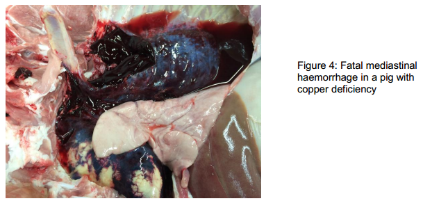
An unusual diagnosis of copper deficiency was diagnosed in a small herd of four-month-old pigs. Two were found dead and post-mortem examination was undertaken at the fallen stock premises in the North of England where an EBLEX-funded initiative is in place. Fatal haemorrhage into the mediastinum and pericardium was found as illustrated in Figure 4 and the vet recalled previous reports in the literature of this pathology due to copper deficiency (Steenmetz and others, 2004, Pig Journal volume 53).
As in the past case, the pigs were being reared on a dairy farm on a predominantly milk-based diet. Profound copper deficiency was confirmed when the livers were submitted (concentrations were <87μmol -er kg DM; reference range 300-5,000) and, perhaps not surprisingly, one pig was also iron-deficient. Cases like this highlight the need to assess the diets of rapidly-growing pigs fed home-mix rations to ensure they are suitable.
Two incidents of PRRS with mortality due to bacterial endocarditis
Interestingly, PRRS was diagnosed at Bury St Edmunds in two further outbreaks of disease with mortality in pigs not vaccinated for PRRS and, in both cases, submitted pigs were found to have bacterial valvular endocarditis. This emphasises the need to consider underlying viral and other disease even when the cause of death is evident.
In the first incident, PRRS and streptococcal endocarditis were diagnosed in finishers from which samples were submitted from on-farm post mortem examinations to investigate the cause of increased postweaning mortality (from three to six per cent), coughing, general poor growth and increased unevenness. Pigs had been seen with purple ears and four of five dead pigs examined on-farm had valvular endocarditis lesions with one dying from severe gastric ulceration and haemorrhage. PRRS virus was detected in serum. Klebsiella pneumoniae subsp. pneumoniae was isolated in mixed growth from pericardium and joint swabs, raising concern that it was involved in a new presentation of disease; however this concern was allayed when two more pigs with valvular endocarditis were submitted to the laboratory for post mortem examination and an untypeable Streptococcus suis was isolated from the heart valves of both pigs. K. pneumoniae is an environmental organism and can contaminate samples collected in conditions where it is difficult to be aseptic, as on farm.
The second outbreak was diagnosed when three sudden deaths from a paddock of 50 13-week-old outdoor pigs prompted submission of two dead pigs. One was in poor body condition and autolysed but post mortem findings pointed to death due to anaemia as a result of haemorrhage from a gastric ulcer. The other pig was in good body condition and had a fibrinous pericarditis and vegetative valvular endocarditis. Actinobacillus equuli was isolated from the heart valve and other internal sites. This organism can cause septicaemia and suppurative conditions in pigs but is not commonly associated with endocarditis which usually involves streptococci or Erysipelothrix species and the concurrent presence of PRRS in this pig may well have predisposed to this presentation. The Actinobacillus equuli was sensitive to all antimicrobials tested.
Septicaemia and endocarditis due to Streptococcus dysgalactiae subsp. equisimilis
Two diagnoses of heart pathology due to Streptococcus dysgalactiae subsp. equisimilis were made on separate unit. Swabs were submitted to Shrewsbury from endocardium and pericardium of a 10-month-old fattening pig that had died suddenly in a good body condition. A pericarditis with marked haemorrhage was found on post-mortem examination. A very heavy pure growth of Streptococcus dysgalactiae was isolated, suggesting streptococcal septicaemia due to this organism as the likely cause of the lesions and death.
In the other case, heart was submitted to Starcross from a 14-week-old pig that was euthanased following a period of malaise. On-farm post-mortem examination revealed a pericarditis and vegetative endocarditis lesions on the mitral valve. Two streptococcal types were obtained; a Group L Streptococcus and Streptococcus dysgalactiae ssp equisimilis. The latter has been isolated from suppurative lesions and septicaemia cases previously in pigs and may have been the significant isolate. The most common causes of bacterial endocarditis in pigs are erysipelas and Streptococcus suis.


