

Emerging Threats Quarterly Report – Pig Diseases - April-June 2014
Among the health issues featured in this quarterly report from the Animal Health Veterinary Laboratories Agency (AHVLA) are vitamin D toxicity in sows, a rare birth deformity in piglets and an increase in the rate of E. coli diagnosis, as well as an update on swine coronaviruses in North America.Highlights
- Update on Swine Coronaviruses in North America
- Vitamin D toxicity incident causing polydipsia in sows
- Rare type of deformities in piglets due to insult in early pregnancy
- Higher diagnostic rate of Escherichia coli disease continues.
New and Emerging Diseases
Analysis of diagnostic submissions from which no diagnosis was made
This report reviews VIDA data where a diagnosis was not reached (DNR) despite the sample receiving “reasonable” testing. This allows monitoring of this class with the aim of providing information on potential new or emerging diseases or syndromes. ‘Prior years’ refers to pooled data for 2009-2013 for GB VIDA data.
DNR by presenting sign and syndrome
A total of 22.1 per cent of GB pig submissions to Q2, 2014 did not reach a diagnosis. This was not significantly increased compared to the overall DNR for the same period in prior years of 18.4 per cent.
Overall DNR rates for SACCVS (20.8 per cent) and AHVLA (22.5 per cent) were not significantly elevated for this period compared to the same period in prior years.
The DNR for GB and AHVLA submissions with a presenting sign of “Wasting” was significantly increased to 38.5 per cent (10/26) compared to 13 per cent in prior years for GB and 38.1 per cent (8/21) compared to 12.62 per cent in prior years for AHVLA. This is reviewed below. DNR for this presenting sign for SACCVS was not significantly increased.
The DNR for GB and AHVLA submissions in “Systemic and Miscellaneous Syndrome” was significantly increased from 8.7 per cent in prior years to 14.8 per cent (20/135) to Q2, 2014 for GB and from 10 per cent to 16.2 per cent for AHVLA (18/111). This is reviewed below. DNR for this syndrome for SACCVS was not significantly increased.
The DNR of 25.7 per cent (19/74) for GB submissions with a presenting sign of ‘Diarrhoea’ was not significantly higher than the DNR of 20.8 per cent in prior years. However, for enteric syndrome GB submissions to Q2, 2014, DNR was 27.5 per cent (30/109) which was significantly increased from 18.85 per cent of prior years. Enteric syndrome DNR was significantly higher for SACCVS submissions (8/31) but not for AHVLA ones (22/78) compared to prior years although the actual DNR was similar for both AHVLA and SACCVS (26-28 per cent). Details of the undiagnosed enteric SACCVS submissions are being reviewed but may well reflect the fact that six of the eight undiagnosed submissions were non carcase (mainly faeces) allowing less comprehensive testing than when pigs are submitted. Undiagnosed AHVLA submissions from pigs with diarrhoea are being tested for porcine epidemic virus as a precaution and none has been detected.
Although there were no overall significant changes in DNR for the following presenting signs, there were alerts generated for certain parameters indicating significant increases in DNR for:
- submissions with a presenting sign of 'Diarrhoea' in Eastern England (10/28)
- submissions from housed pigs 'Found Dead' (10/38)
- carcass submissions with a presenting sign of 'Respiratory' (4/19)
The relevant submissions in each case were reviewed to investigate whether there were common features or causes for concern. One factor which may be influencing some submissions (found dead and diarrhoea) is enteric disease due to Escherichia coli which is detailed in this report under the section on “Changes in disease patterns and risk factors”. Otherwise there was no evidence for a new and emerging syndrome in the undiagnosed submissions.
There was no other overall significant increases in DNR for other syndromes or presenting signs for GB, AHVLA or SACCVS data.
DNR for submissions to AHVLA with a presenting sign of 'Wasting'
The DNR for this presenting sign was also significantly increased in the first quarter of 2014 and the undiagnosed AHVLA submissions were reviewed in the last 'Emerging Threats' report and did not indicate a common clinical or pathological condition. The undiagnosed submissions in April to June were reviewed. Two were non-carcase submissions limiting full diagnostic examination, one was of pigs in an inappropriate disease phase and the fourth was of dead weaners with anaemia and necrotic enterocolitis the cause of which was not diagnosed but histopathology indicated a bacterial enteritis.
Overall, these undiagnosed cases did not suggest a new and emerging disease, or the presence of periweaning failure to thrive syndrome (see www.defra.gov.uk/ahvla-en/files/pub-vet-pfts.pdf).
DNR for Systemic and Miscellaneous submissions to AHVLA
The DNR for this syndrome was also significantly increased in the first quarter of 2014 and the undiagnosed AHVLA submissions were reviewed in the last 'Emerging Threats' report and did not indicate a common clinical or pathological condition.
The undiagnosed submissions in April to June were reviewed. All but one were carcass submissions with a range of pathologies (enteric, polyserositis, pneumonia) with the causes not identified, in some due to prior treatment, in others because the disease phase was inappropriate and in two there was no clear reason for the lack of diagnosis. This will be kept under review but overall, there are not consistent findings at this stage to suggest an emerging syndrome.
Analysis of undiagnosed submissions to Q2, 2014 has not revealed evidence of a new and emerging syndrome in GB pigs.
Update on Swine Coronaviruses in North America
As described in previous quarterly reports, porcine epidemic diarrhoea (PED) emerged in 2013 in a virulent form in the United States of America (US) and spread to Canada in January 2014. Japan also reported outbreaks from October 2013 and has continued to have outbreaks since; a four per cent reduction in slaughter pig numbers is predicted for the last six months of 2014.
The disease in the US was made reportable by USDA and by July 2014, 30 US States had experienced at least one outbreak. Since then, USDA working with veterinarians and the pig industry have defined different infection statuses (suspect, presumptive positive and confirmed positive) together with a requirement for specific actions to be undertaken at each stage. In Canada, the spread of infection has been more limited, aided by the very stringent biosecurity initiatives on infected farms, vehicles and at collection centres and abattoirs, and the aim is to eliminate infection from Canada by autumn 2014.
In both countries, the rate of spread has fallen during summer months as weather conditions reduce virus survival and cleaning and disinfection is often more effective. Recent work (Vlasova and others, 2014) has shown that there is complex and rapid evolution of PEDv strains in the US.
These strains differ from the CV777 virus from Europe and from virus from a pig in a PED outbreak in England in 2000 but there is very limited virus material available from some parts of the world, including Europe on which to base a comparison.
The experiences of the disease in North America are helping to inform contingency plans and discussions on how PEDv may be handled should it enter GB pigs.
The Pig Health and Welfare Council (PHWC) surveillance subgroup is leading actions resulting from the exotic and emerging diseases roundtable (www.bpex.org.uk/R-and-D/Pig-Health/diseases.aspx) in which AHVLA is particularly involved in horizon scanning and early detection initiatives.
PED PCR remains the test of choice for diagnosis. PEDv PCR testing of routine diagnostic samples submitted to AHVLA from pigs with diarrhoea has continued through Defra-funded surveillance and no PCR-positive samples have been detected.
Enhanced surveillance funded by BPEX was recently introduced whereby samples from outbreaks of diarrhoea in pigs, which would not otherwise have been sent to AHVLA, can be submitted for PEDv PCR free of charge.
Swine deltacoronavirus has been detected in US and Canada and testing guidelines for this virus and PEDv have been included in the National Pig Association’s voluntary protocol for non-statutory pathogens in live imported pigs. No PEDv has been detected through current surveillance in GB pigs.
Maintaining surveillance and implementation of the exotic and roundtable recommendations remain key activities for all stakeholders. Funding for diagnosis of suspected virulent PED outbreaks, should they occur, remains available from the Defra-funded pig scanning surveillance project (ED1200).
Vitamin D toxicity incident causing polydipsia in sows
All sows on two related breeding units were affected by hypervitaminosis D as a result of Vitamin D being added in significant excess to the vitamin premix used to prepare the affected ration. Fortunately, the premix was only used in feed for these two pig units.
Clinical signs were noticed from 14 to 20 days after the affected feed batches were first fed and initially were of marked polydipsia and secondary polyuria, followed soon afterwards by lethargy, inappetence and weight loss.
Post-mortem findings were largely unremarkable apart from mottling and streaking of the kidneys (Figure 1), biochemistry revealed consistently elevated serum calcium concentrations and histopathology showed evidence of a sub-acute nephropathy with calcium phosphate mineralisation (Figure 2).
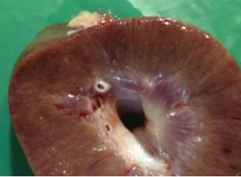
(Image from Rudolf Reichel, AHVLA)
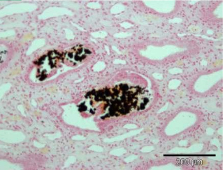
(Image from Martin Jeffrey, AHVLA)
The affected feed was replaced immediately concerns arose and, while investigations were in progress, voluntary restriction of cull sows on-farm for a specified period was agreed to protect the food chain.
The attending veterinary surgeon raised the possibility of vitamin D toxicity after determining that the vitamin-mineral premix batch had only been used for the affected units and other feed ingredients had been included in other rations without incident. This suspicion was consistent with the clinical and laboratory findings and was confirmed when markedly elevated vitamin D concentrations were found in sera sent to an NHS laboratory for testing and in feed samples.
The clinical signs took almost a month to abate after withdrawal of the feed, which may reflect the long half-life of 25/OH Vitamin D3. There was significant weight loss, necessitating culling of some sows, especially in those which had been lactating and thus exposed to a greater vitamin D intake. There were also secondary reproductive effects.
This incident exemplifies the collaborative approach to an unexplained disease outbreak reported to AHVLA by a veterinary practitioner whose on-farm observations triggered complementary laboratory investigations and achieved a diagnosis. The case will be presented at the Pig Veterinary Society autumn meeting in November highlighting aspects relating to investigating suspected feed-related disease with potential food safety issues.
Ongoing Investigations
Klebsiella pneumoniae septicaemia
Each summer since 2011, outbreaks of septicaemia in piglets due Klebsiella pneumoniae subsp. pneumoniae (Kpp) have been diagnosed on units in East Anglia.
The principal features of the outbreaks diagnosed so far were:
- Sudden or rapid deaths of pre-weaned pigs in good body condition
- Piglets affected from about two-weeks-old to weaning
- Only diagnosed in weaners on one occasion 36 to 48 hours after being weaned
- Outbreaks diagnosed in summer months (May to September) each year since 2011
- All but one outbreak in outdoor herds
- All outbreaks in East Anglia
The Pig Expert Group has also received reports of recent outbreaks in other countries where, like here, Kpp previously caused septicaemia only sporadically in individual pigs. Postmortem examination and laboratory testing are necessary to confirm a diagnosis and distinguish Kpp from other causes of septicaemia in pre-weaned pigs such as Streptococcus suis, Actinobacillus suis, Haemophilus parasuis and erysipelas.
All the AHVLA outbreak isolates have been the same sequence type (MLST 25) with a particular combination of a small plasmid and a particular virulence gene not present in non-outbreak isolates. There has not been significant antimicrobial resistance in outbreak isolates but ampicillin/amoxicillin resistance is innate for Kpp.
No outbreaks were diagnosed in GB between April and June 2014, however, in early July, AHVLA Starcross diagnosed the first outbreak to occur outside East Anglia, details of this will be included in the July to September 2014 'Emerging Threats' report and an alert was sent to veterinarians.
Further information on Kpp septicaemia is available on this link: www.defra.gov.uk/ahvla-en/publication/pig-survreports.
Unusual Diagnoses and Presentations
There were a number of unusual diagnoses this quarter; details of these have been included in monthly AHVLA or SACCVS reports, www.defra.gov.uk/ahvla-en/publication/pig-survreports-monthly/. These will be kept under review to assess whether they justify initiation of emerging disease investigations.
Rare type of deformities in piglets due to an undetermined insult early in pregnancy
A few piglets with unusual craniofacial deformities were born at term in litters from any parity of sow on an indoor herd in which other piglets in the litters were unaffected.
Arhinia was present accompanied by absence of the nasal cavity and olfactory lobes (Figure 3). The defects were either bilateral or unilateral; in humans, this is a rare form of deformity.
Based on the timing of embryonic development and the fact that olfactory pits are present in 7-mm pig embryos (about 20 days), the histopathologist estimated that any insult leading to arhinia would have been before 20 days gestation. Thus there was a tight window for causing these lesions and, as disease only occurred over a six-week period, it was transient, although not a point exposure as sequential weekly farrowing batches had a few affected piglets.
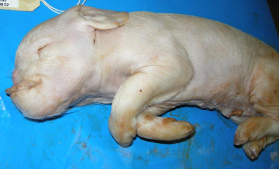
(Image from Julie Wessels, AHVLA)
Piglets were delivered full-term at the end of May so exposure to the insult would have been early February. The sows were liquid-fed on a wide variety of food by-products from multiple sources which are added to a wheat, barley and soya mix for feeding. There was no evidence of infectious exposure; and an association between this type of craniofacial malformation and viral infection was considered unlikely.
It is possible that the malformations were a consequence of complex interaction between maternal and environmental factors. Involvement of toxic plants was very unlikely as the sows were indoors. The submission of typical cases in incidents of this nature is very useful in characterising the deformities, exploring possible aetiologies and allowing assessment of the likely timing of any insult.
If further cases are encountered, control material will also be collected from unaffected age-matched piglets and epidemiological details will be collated of any exposures of sows to possible toxins or other insults, to add to the information from this case.
Systemic porcine cytomegalovirus infection in fading weaners
Porcine cytomegalovirus (PCMV) is occasionally diagnosed in AHVLA, usually associated with rhinitis, and sometimes wasting, post-weaning.
An uncommon systemic manifestation of PMCV was diagnosed with swine influenza and streptococcal disease in a submission of live piglets, at the point of weaning, to Thirsk to investigate a problem of fading starting about later post-weaning affecting five to seven per cent of pigs. The pigs had significant mucoid rhinitis (see Figures 4 and 5), pneumonias of varying severity and two also had pleuritis.
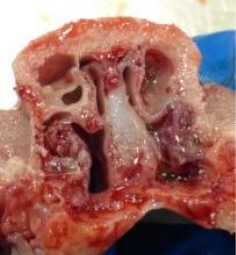
(Image from Rudolf Reichel, AHVLA)
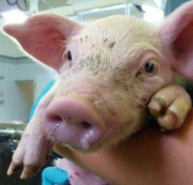
(Image from Rudolf Reichel, AHVLA)
Swine influenza (pandemic H1N1 2009 strain) and Streptococcus suis type 8 were detected in the lungs and histopathology was consistent with swine influenza with a secondary bacterial infection.
Histopathology revealed that the severe rhinitis was due to PCMV infection and, unusually, lesions in the kidneys pointed to systemic PCMV infection in three piglets. It was likely that this early viral disease in piglets predisposed to the wasting seen later on the unit.
Atrophic rhinitis in a previously unaffected Scottish pig herd
Atrophic rhinitis was diagnosed by SACCVS in post-weaned pigs on an indoor breeding to finishing unit.
Signs of respiratory disease, characterised by sneezing and nasal discharges, developed post-weaning with uneven growth rates and ill thrift but minimal mortality in affected pigs.
Two six-week-old pigs in poor body condition were euthanased and partial atrophy of the turbinates was found on one side of the nasal chamber.
Atrophic rhinitis is now rare in GB pig herds with the last VIDA diagnosis being recorded in 2010; the diagnosis prompted further investigation to determine the source of infection.
Changes in Disease Patterns and Risk Factors
Higher diagnostic rate of Escherichia coli disease continues
In the last 'Emerging Threats' report, it was reported that GB diagnoses of disease incidents due to Escherichia coli had shown an increased trend in the last three years and this trend continued in this quarter despite reduced submissions. The number of actual incidents diagnosed in this quarter was nine; five enteric, three oedema disease and one colisepticaemia, however expressed as a percentage of diagnosable submissions, bearing in mind that there was a reduction in submissions this quarter, this equates to 6.2 per cent diagnosed between April and June 2014 which is the highest for this quarter of the year since 2006.
Alongside this higher diagnostic rate, AHVLA have diagnosed a few outbreaks of enteric colibacillosis in slightly older post-weaned pigs over recent months. The pigs have been dehydrated with fluid enteropathies and two examples of confirmed enteric colibacillosis are illustrated in Figures 6 and 7 below. A number of different E. coli serotypes have been involved in the enteric cases including O149:K981,K88ac (Abbotstown), O139:K82 (E4), O157: K’V17’ V17, and O138:K81 (E57).
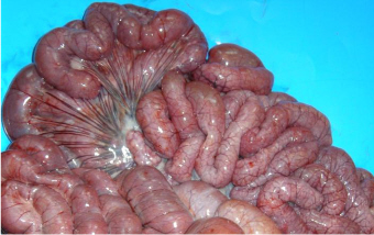
(Image from Susanna Williamson, AHVLA)
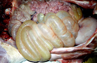
(Image from Susanna Williamson, AHVLA)
In response to this observation, those undertaking post-mortem examinations have been advised to initiate culture for E. coli if the gross lesions are suggestive of this disease even if the pigs are older than those in which enteric colibacillosis is usually expected. In addition, it is worth remembering that enteropathogenic E. coli in pigs are not always haemolytic or K88 (F4) antigen positive and that serotyping of non-haemolytic, K88 antigen negative E. coli is merited if clinical history and gross lesions are suggestive of enteric colibacillosis and other enteropathogens are not detected.
Information was circulated to Veterinary Investigation Officers to raise their awareness of these observations. A funding proposal is being submitted which will include genetic analysis of E. coli isolates from pigs stored at AHVLA and, if successful, the work will help determine whether there is any change in virulence or other genes in more recent isolates.
Fewer swine dysentery diagnoses in GB pigs from April to June 2014
The percentage of tested submissions diagnosed by AHVLA and SACCVS as swine dysentery fell to 1.9 per cent (two diagnoses) in this quarter compared to 5.6 per cent (11 diagnoses) in the first quarter of 2014 as illustrated in Figure 8.

(Note data not yet recorded for Q3, 2014)
One of the Q2 diagnoses was made in the Thirsk region in a small pig herd in growing pigs with diarrhoea, the other was notable in that it was the first diagnosis in the East Anglian region since 2012. AHVLA made no diagnoses of swine dysentery in East Anglia in 2013 and only one, in smallholder pigs, in 2012.
The East Anglian Brachyspira hyodysenteriae isolate was sensitive to tiamulin but the East Anglian producer involved took the decision to depopulate and clean and disinfect to eliminate infection from the unit. Whether this reduction in diagnostic rate reflects a true reduction in disease is uncertain and diagnoses will be monitored. It is possible that good weather conditions have assisted those implementing control programmes.
Swine dysentery remains one of the priority diseases targeted for control by the GB pig industry and more information is available on swine dysentery from BPEX on the following link: www.bpex.org.uk/2TS/health/swinedysentery.aspx.
Antimicrobial minimum inhibitory concentrations in UK Mycoplasma hyopneumoniae
There is no routine GB surveillance for antimicrobial sensitivity in Mycoplasma hyopneumoniae, in part due to the fact that culture to obtain isolates is rarely undertaken as PCR is used for diagnosis.
M. hyopneumoniae is one of the respiratory pathogens involved in porcine respiratory disease and, although vaccination is widespread, it is sometimes found to be playing a role in disease outbreaks, often concurrent with other respiratory pathogens.
The Defra-funded pig scanning surveillance project (ED1200) contributed funding to undertake minimum inhibitory concentration (MIC) assays for 13 antimicrobials on UK M. hyopneumoniae isolates provided by Andrew Rycroft at the Royal Veterinary College on which whole genome sequencing had already been performed. This was considered a useful preliminary step to investigate possible antimicrobial resistance in M. hyopneumoniae isolates.
Preliminary results were presented in a poster (Ayling and others, 2014) and some UK isolates were found to have higher MIC values to certain antimicrobials. No MIC breakpoints exist to relate the values found to efficacy in vivo, however, further investigation is in progress at the RVC including genome analysis to detect resistance genes.
Pasteurella multocida typing from septicaemia cases in pigs
A presentation at the 2013 ESPHM from Spain (Cardoso-Toset and others, 2013) reported involvement of a multilocus sequence type (ST47) of Pasteurella multocida in septicaemic disease in pigs.
In response to a small cluster of cases of P. multocida septicaemia diagnosed in pigs by AHVLA in 2013, multilocus sequence typing (MLST) was undertaken on the isolates available from pigs from 2011-2013, including some which caused septicaemia.
Four different STs were identified (3, 10, 11 and 12), and, although the existing database of typed isolates is fairly limited (mainly from UK and Spain), all those identified had previously been detected in England (and Wales) from a variety of hosts. Although the two most recent septicaemia outbreaks diagnosed at Bury St Edmunds were due to ST11, there was no evidence of either ST47 or of a novel or single emergent strain associated with systemic P. multocida infection in pigs.
This provides useful background information on archived P. multocida isolates should there be an upsurge in pasteurellosis in the future.
Surveillance for non-statutory pathogens in culled wild boar in the Forest of Dean
BPEX funded AHVLA to undertake surveillance of wild boar culled over autumn and winter of 2013-14 for selected non-statutory pathogens in an established population in the Forest of Dean.
The following is a summary of the surveillance findings:
- Faeces and serum samples collected from 91 wild boar culled from the Forest of Dean were tested for non-statutory endemic pathogens of GB pigs.
- Serological evidence of exposure to two Leptospira serovars was detected with a combined seroprevalence of nearly 18 per cent.
- Leptospira Bratislava, the Leptospira serovar to which most seropositive wild boar had antibody, is pig-adapted and also occurs in a range of wildlife species. The seroprevalence detected was significantly higher in older pigs, and the results are consistent with endemic infection in the wild boar population.
- A lower seroprevalence was detected to Leptospira Javanica which is reported in the literature to be associated with a rodent reservoir and is zoonotic.
- One adult female was positive for antibody to porcine reproductive and respiratory syndrome virus by ELISA but active infection was not detected, indicating potential for infection but not clear evidence that the wild boar population is a reservoir of infection to domestic pigs.
- Infection with, or exposure to, other pathogens tested was not confirmed and with the given population and sample size this indicates at least 95 per cent confidence that the prevalence of those pathogens in this wild boar population is less than five per cent.
- No Salmonella species were detected, culture results for a proportion of faecal samples may have been affected by the time interval between collection and culture.
These results are relevant for a long established wild boar population in a forested region of England which has a relatively low commercial pig density and should not be extrapolated to wild boar populations which exist, or could establish, in other regions.
Suggestions regarding future wild boar surveillance were included in the full report which will be made available by BPEX (Williamson, S., Smith, R. and Barlow, B. 2014. Surveillance for Non-Statutory Pathogens In Culled Wild Boar in the Forest of Dean. BPEX-Funded Project Report RDOR1035).
Horizon Scanning
African Swine Fever spread in EU in Eastern Europe
In May, Poland reported further African Swine Fever (ASF) in wild boar and then in June 2014, Latvia joined Poland and Lithuania as the third EU member state to report disease in wild boar but also reported an outbreak in backyard pigs. Both were in an area which was already restricted due to ongoing classical swine fever (CSF) and incursion is likely to have occurred through movement of infected wild boar from Belarus. Further spread has occurred since including into commercial pigs in Lithuania in July.
Preliminary outbreak assessments are available on this link: www.defra.gov.uk/animal-diseases/monitoring/poa/.
This spread underlines the importance of the activities in relation to the PHWC exotic and emerging diseases roundtable recommendations (www.bpex.org.uk/R-and-D/Pig-Health/diseases.aspx) to help mitigate the risk of incursion and increase awareness of notifiable diseases including ASF amongst pig keepers and veterinary surgeons attending pigs.
The Pig Veterinary Society is holding a CPD event which will include ASF and CSF to increase awareness in practitioners (both pig specialist and non-specialist).
Brachyspira hampsonii reported in Belgian pigs
Infection with Brachyspira hampsonii, a new potentially pathogenic Brachyspira species described in pigs in 2012 in North America, was reported in two gilts, imported into Belgium from the Czech Republic, examined during quarantine (Mahu and others, 2014).
The pigs had ill-thrift and dilated large intestines containing soft to watery non-haemorrhagic contents. Bird transmission has been implicated in transmission of infection elsewhere in the world.
B. hampsonii has not been detected in GB pigs. No specific surveillance in place for this organism but AHVLA and SACCVS have the methods to identify it in diagnostic submissions.
Livestock Associated-MRSA in Australia and Northern Ireland
Livestock-associated methicillin-resistant Staphylococcus aureus (LA-MRSA) ST398 was reported in pigs in Australia (Groves and others, 2014).
The authors believe infection was most likely to have been introduced by transmission into pigs from an infected human or other livestock species.
In July, the first detection of LA-MRSA in a pig in UK was announced in a letter to the Veterinary Record (Hartley and others, 2014). The LA-MRSA was detected in Northern Ireland in a single pig as an incidental finding during diagnostic investigation of respiratory disease due to porcine reproductive and respiratory syndrome virus. Veterinary authorities in Northern Ireland (DARDNI) are leading the investigation.
No MRSA was detected in UK breeding pig herds sampled during an EU survey in 2008 which detected LA-MRSA in 12 of the 24 member states reporting results (EFSA, 2009). Analyses of the results of this survey at country-level demonstrated a strong positive association between the prevalence of MRSA-positive breeding holdings and MRSA-positive production holdings, suggesting a vertical dissemination of MRSA between the holdings, with transmission in breeding pigs being a potential route (EFSA, 2010). There has been no more recent specific surveillance for LA-MRSA in UK pigs.
Aujeszky’s disease in vaccinated pigs in China
Outbreaks of Aujeszky’s disease (AD) have been reported in ADV vaccinated pigs in China (Tong-Qing An and others, 2013) and involving a virus variant. There are no semen or live pig imports from affected areas and this has not been reported in European herds. This is noted as a point of information for GB rather than a current threat.
Pig movements in Scotland
Horizon scanning includes consideration of factors affecting the risk of threats occurring and identifying changes in these factors.
Two such factors are the pattern of pig movements and the degree of interaction between commercial and non-commercial pig sectors and an analysis of both in Scotland was recently published (Porphyre and others, 2014).
This analysis helps inform biosecurity and surveillance initiatives by identifying features affecting the risk of disease spread using data from pig movements made over a 15-month period to May 2013.
Further Reading
You can view the full report by clicking here.
Find out more about the diseases mentioned by clicking here.
September 2014


