


3.2 How does Lawsonia intracellularis induce the immune response?
Information has recently become available about the humoral immune response in pigs naturally or experimentally infected with L.intracellularis. In one study, IgG could be first detected two weeks after challenge of 5-week-old pigs with pure culture of L.intracellularis. Antibody levels (IgG) peaked around the end of the third week and then tended to drop (Knittel et al. 1998).
A unique study (Collins et al. 2001) indicated development of immunity in pigs after re-inoculation. Those animals were challenged a second time after fecal shedding of L.intracellularis had ceased to be detected. Re-inoculated pigs were evaluated clinically and the feces tested by PCR to detect any shedding. Animals previously infected did not shed detectable numbers of L.intracellularis in their feces and had no clinical signs of the disease. Those findings suggested that bacteria used in the second challenge were probably inactivated before entry and colonization of mucosal cells.
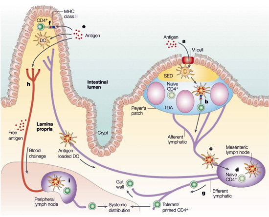
Figure 3.2 a Antigen uptake and recognition by CD4+ T cells in the intestine (Mouse model)
Antigen might enter through the microfold (M) cells in the follicle-associated epithelium (FAE) (a), and after transfer to local dendritic cells (DCs), might then be presented directly to T cells in the Peyer’s patch (b). Alternatively, antigen or antigen-loaded DCs from the Peyer’s patch might gain access to draining lymph (c), with subsequent T-cell recognition in the mesenteric lymph nodes (MLNs) (d). A similar process of antigen or antigen-presenting cell (APC) dissemination to MLNs might occur if antigen enters through the epithelium covering the villus lamina propria (e), but in this case, there is the further possibility that MHC class II+ enterocytes might act as local APCs (f). In all cases, the antigen-responsive CD4+ T cells acquire expression of the α47 integrin and the chemokine receptor CCR9, leave the MLN in the efferent lymph (g) and after entering the bloodstream through the thoracic duct, exit into the mucosa through vessels in the lamina propria. T cells which have recognized antigen first in the MLN might also disseminate from the bloodstream throughout the peripheral immune system. Antigen might also gain direct access to the bloodstream from the gut (h) and interact with T cells in peripheral lymphoid tissues (i). Mowat/Nature Reviews – Immunology Vol. 3, April 2003, 331-341
Cell-mediated immune response is an important feature involved in infection caused by intracellular organisms (Goldsby et al. 2000). Descriptive immunocytological studies of intestinal tissue sections of pigs affected by both clinical forms of proliferative enteropathy have shown a mild infiltration of cytotoxic Tcells, macrophages and B lymphocytes carrying MHC class II structure at the beginning of the cell-mediated immune response (McOrist et al. 1992). Mucosal local humoral immunity, represented by secreted IgA, is a known relevant defense mechanism against enteropathogenic microorganisms (Lamm et al. 1995, Goldsby et al. 2000). Immunohistochemistry studies of intestinal sections of pigs naturally infected by proliferative enteropathy demonstrated a large accumulation of IgA in the apical cytoplasm of proliferating enterocytes (Lawson et al. 1979, McOrist et al. 1992). A systemic cell-mediated immune response was successfully detected in PBMC’s using an assay for specifically-produced interferon gamma (Guedes and Gebhart, 2003) and IgA could be detected in intestinal lavages of challenged pigs (Guedes 2002). The roles of systemic and local cell-mediated immune responses and local production of IgA specific to L.intracellularis infection have not yet been studied.
Recent studies from Guedes and Gebhart (2003) and from MacIntyre, Smith, Shaw, Thomson and Rhind (2003) have thrown some light on the cellular immunity related to Lawsonia intracellularis.
From the results the authors concluded that a depression of T-cells, especially the CD3+ and CD8+ in the villus epithelium, was a reflection of the inability of the host to mount a cellular response to the intracellular pathogen Lawsonia intracellularis. This resulted in the hypothesis of a down regulation of the ligand for intraepithe-lial lymphocytes associated with the disease.
Macrophages were present in larger numbers in crypts showing hyperplastic lesions as compared to those infected but not hyperplastic. This massive infiltration of activated macrophages, peaking on day 14, may be the key factor in the development of the haemorrhagic form of the disease. (Figure 3.2 b)
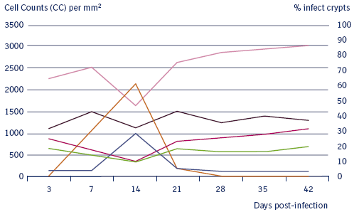
Figure 3.2 b
Immune response to a Lawsonia intracellularis
challenge.
MacIntyre, Smith, Shaw, Thomson and Rhind / Vet. Path.
40:4 (2003).
The reduction of B-cells is unexpected given the accumulation of IgA within the enterocytes. A phenomenon which has been seen by others. An explanation for this could not be given.
The onset and duration of systemic cell-mediated and humoral immune responses and fecal shedding in pigs experimentally infected with pure culture of L.intracellularis was accessed (Guedes and Gebhart 2003) (Figure 3.2c). Humoral and cell-mediated immune responses were initially detected two weeks after exposure in pigs challenged with the pathogenic isolate. Humoral and cell-mediated immune responses were still detected in some pigs 13 weeks after exposure. Fecal shedding was initially detected one week and lasted, intermittently, 12 weeks post exposure in challenged pigs. Similarly, in experimental infections of mice, interferon-gamma was found to play a role in limiting intracellular infection and increased cellular proliferation associated with L.intracellularis (Smith, et al. 2000). Thus, animals exposed to a pathogenic pure culture isolate of L.intracellularis demonstrated long-term shedding of and specific immune responses to the organism.
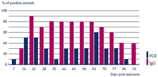
Figure 3.2 c
Induction of seroconversion
and shedding
after Lawsonia intracellularis infection.
PCR and IPMA results for serum IgG against Lawsonia intracellularis
in pigs.
Guedes and Gebhart/ Vet. Microb. 91 (2003) 135–145.
Results from vaccination studies with Enterisol® Ileitis show little reaction in the IFA and IPMA tests on serum IgG.
The response of the immunological system to an intracellular infection as caused by Lawsonia intracellularis is still poorly understood and will be a major field for investigation in the future.
© Boehringer Ingelheim Animal Health GmbH, 2006
All rights reserved. No part of this Technical Manual 3.0 may be reproduced or transmitted in any form or by any means, electronic or photocopy, without permission in writing from Boehringer Ingelheim Animal Health GmbH.