


The reproductive physiology and anatomy of the sow
Learn about the sow's reproductive system to create successful breeding 22 February 2023
 8 minute read
 By:
By: Editor's note: This article is part of the Purdue University Pork Archive.
An understanding of the sow's reproductive system is essential for a successful mating program, whether AI or natural service is used. This discussion will be divided into three areas:
- Anatomy of the reproductive tract
- Estrous cycle, estrus and ovulation
- Embryonic and fetal development
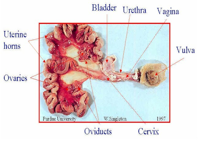
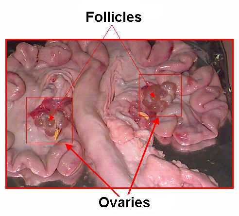
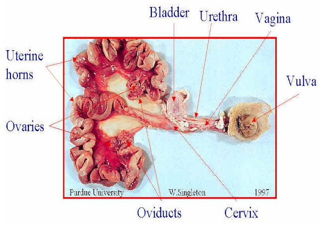
The primary structures of the female reproductive tract are the ovaries, they have two major functions:

(1) to produce ova, the female germ cells and
(2) to produce the hormones progesterone and estrogen.
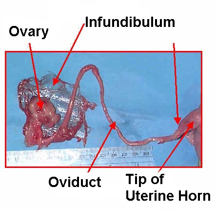
Each ovary is surrounded by a thin membrane called the infundibulum which acts as a funnel to collect ova and divert them to the oviduct. The oviduct is about 6-10 inches long and acts as the site of fertilization.

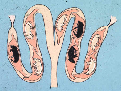
There are two uterine horns. Each is 2-3 feet in length in the non-pregnant sow. They act as a passageway for sperm to reach the oviduct and are the site of fetal development. The uterine body, which is small compared to some other species, is located at the junction of the two uterine horns.
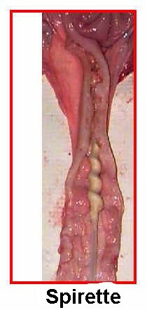
The cervix is a muscular junction between the vagina and uteri. It is the site of semen deposition during natural mating and AI. It is dilated during heat (estrus) but constricted during the remainder of the estrous cycle and during pregnancy.
The vagina extends from the cervix to the vulva and serves as a passageway for urine and the piglets at birth. The bladder is connected to the vagina by the urethra.
The vulva is the external portion of the reproductive tract. It often becomes red and swollen just prior to estrus and this swollen condition is usually more pronounced in gilts than in sows.
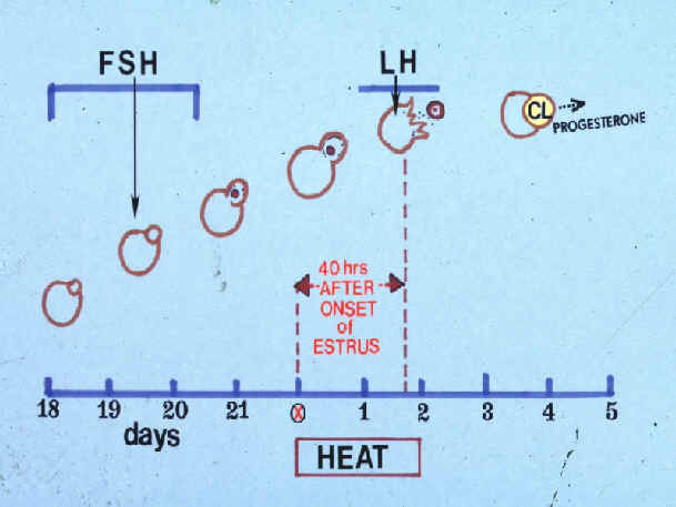
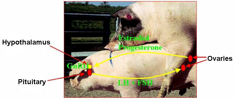
The hypothalamus located at the base of the brain secretes gonadotropin releasing hormone (GnRH) which regulates the anterior pituitary gland to secret FSH (Follicle Stimulating Hormone) and LH (Luteinizing Hormone) into the blood which stimulates the production of the ovarian hormones, estrogen and progesterone, which in turn regulate the reproductive process. Oxytocin is released from the posterior pituitary gland.
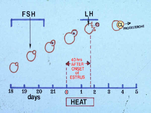
Estrous Cycle, Estrus and Ovulation (see Figure 2)
Non-pregnant and non-lactating sows and gilts display estrus or standing heat on a regular basis throughout the year. The estrous cycle is normally 21 days and is defined as the time between the onset of one estrus to the onset of the next. The cycle length can range from 18-24 days.
Lactation or the nursing stimulus inhibits the estrous cycle and sows will not, as a rule, return to heat until the litter is weaned. Days from weaning until estrus is influenced by such factors as length of lactation, parity, season and nutritional level, but should range from 4 to 7 days.
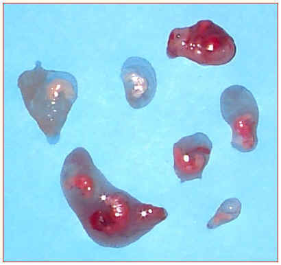
As estrus or heat approaches, 6-10 follicles or "blister like" structures form on each ovary. Follicular growth accelerates about 3 days before estrus and is influenced by FSH or follicle stimulating hormone released from the pituitary gland, located at the base of the brain. A maturing ovum is held within each follicle. Granulosa cells within the follicle secrete estrogen, a hormone, which among other things is responsible for the typical signs of estrus. Ovulation, or release of the ova, is stimulated by LH. Ovulation occurs about 40 hours after the onset of estrus, but this interval is variable.
Several factors can influence ovulation rate or number of ova shed.
- Age. Sows may ovulate 18-20 ova while gilts may ovulate 12-14 ova.
- Nutrition. Flushing (increased energy levels prior to estrus) may increase ovulation rate yet may have little effect on the ultimate litter size.
- Breed. The white or maternal breeds generally have a higher ovulation rate. Crossbred females generally have a higher ovulation rate than either of the parent breeds.
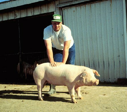
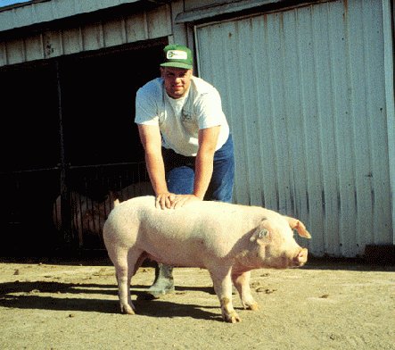
The onset and disappearance of estrus and estrus behavior is gradual and there are individual differences among females (see Fig. 3). The primary sign, and most reliable sign, of estrus is "standing" while another sow or the boar mounts. Many females will stand for the "back pressure test" when applied by the herdsperson. A higher percentage of females will respond to the "back pressure test" if there is a boar present. Therefore, use of an intact or a vasectomized boar is an important part of a regular heat detection program. Boars secrete pheromones (odors) in their salivary glands which elicit the standing reflex of the female. Mature boars are superior to young boars in stimulating this response.
Secondary signs of estrus include:
- Red, swollen vulva which is usually more pronounced in gilts than in sows.
- Increased nervous activity.
- Desire to seek the boar.
- Loss of appetite.
- Male-like sexual behavior (pursuing, nosing and mounting other females).
- Change in vocalization (grunts and growls).
- Increase in vaginal mucous (thumb check).
Sexual Behavior in Swine
Length of estrus or heat is variable and may last only 12 hours in gilts or up to 60 hours or more in sows. Since the actual time of the onset of estrus is rarely known, it is recommended that a female receive at least two matings during estrus. This helps insure that sperm are present at an optimum time relative to ovulation for fertilization to occur.
Inseminate a female about 12 hours after the beginning of standing estrus is observed and again 18 to 24 hours after the first insemination. The optimum time to mate will vary from farm-to-farm. Two services as compared to one may increase conception rate and litter size by approximately 10%.
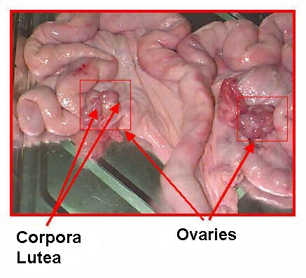
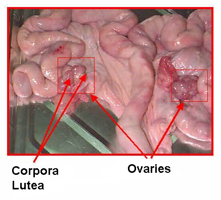
After ovulation, the follicular structures are transformed into corpora lutea. Beginning on about day 1 to 2 after mating, they produce and release the hormone, progesterone which is responsible for maintaining pregnancy. If conception does not occur, prostaglandin (F2a) is secreted by the uterus causing the corpora lutea to regress and cease producing progesterone on about day 17 of the cycle. At this time, a new set of follicles begins to develop, and the process is repeated. If conception does occur, the corpora lutea remain functional and continue to secrete progesterone throughout pregnancy.
Fertilization, embryonic and fetal development
Semen is deposited into the cervix during mating or insemination. Muscular contractions of the cervix stimulate ejaculation of the boar and is the basis of the boar's ejaculatory response to the "gloved hand" or "digital pressure" technique of semen collection.
Thirty to sixty billion sperm are deposited by the boar during natural service or 2 to 6 billion during insemination but only a small fraction of these reach the oviduct and the vicinity of the ova. Even though sperm are motile, they do not propel themselves through the female's tract. Sperm transport results from uterine contractions. These contractions are stimulated by oxytocin released from the pituitary gland. Its release is mediated by the stimuli from mating behavior and copulation. Keep this in mind when inseminating a sow or gilt. Face-to-face contact with a boar, plus tactile stimulus during and after insemination may improve sperm transport. Sperm must reside in the female from 6-10 hours before they are capable of fertilization. This process is called capacitation and it includes both physical and biochemical changes within the sperm.
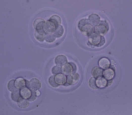
Fertilization, or union of sperm and ova occurs in the upper 1/3 of the oviduct. Fertilization rate is virtually 100% if the female is mated to a fertile boar at the correct time relative to ovulation. About 2 days after fertilization, the blastocysts enter the uterus. They are in the 4-8 cell stage and begin to space themselves equal distances apart in the uterine horns. The blastocysts can migrate from one horn to another during this stage.

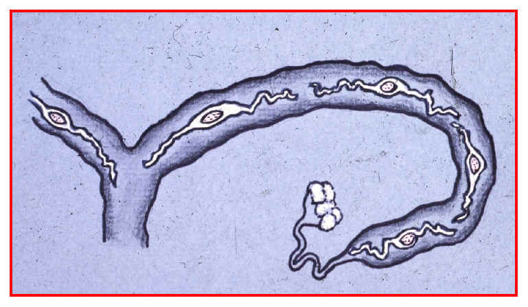
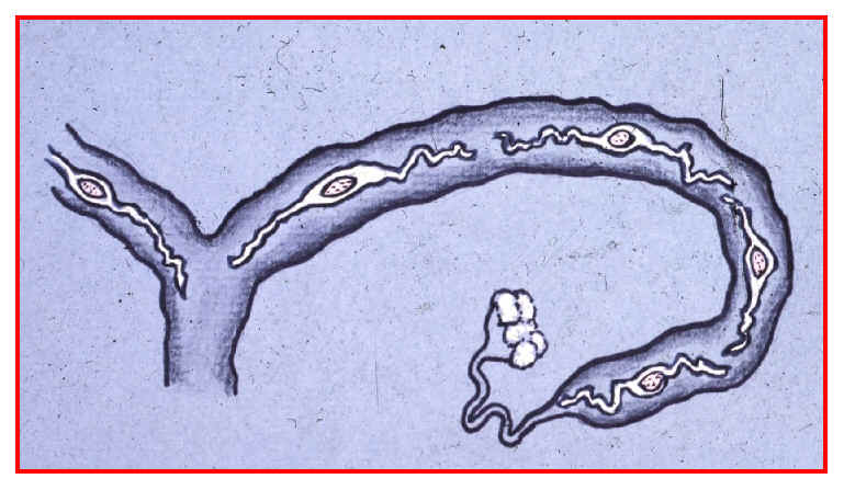
One of the most critical periods of pregnancy is from approximately day 11-16 after mating. The blastocysts, elongate into long (2-3 feet), stringy masses, and they begin to attach to the uterine wall. The greatest potential loss in litter size may occur during this stage of development because of attachment failures. On the average, 17 ova are shed at estrus yet only about 12 are accounted for as blastocysts when attachment complete. There is thought to be some factor(s) which limit the number of blastocysts that can attach in a given uterus. Research has shown that overcrowding is not a major limiting factor. Embryonic survival may be related to certain uterine secretions which have not presently been well defined. Environmental stressors such as high temperatures and fighting as a result of mixing or regrouping animals also adversely influence implantation and embryo survival. Generally, the presence of at least four blastocysts are required in order for pregnancy to continue.
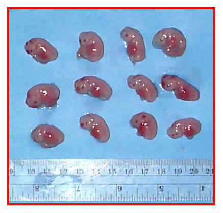

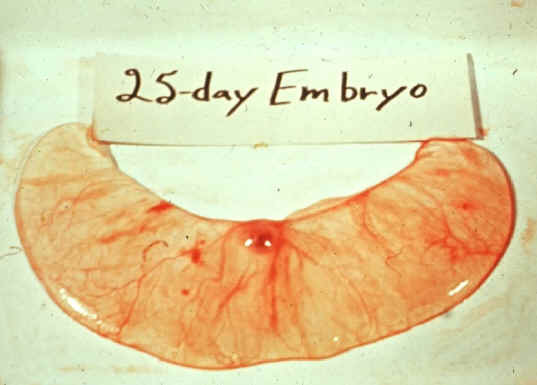
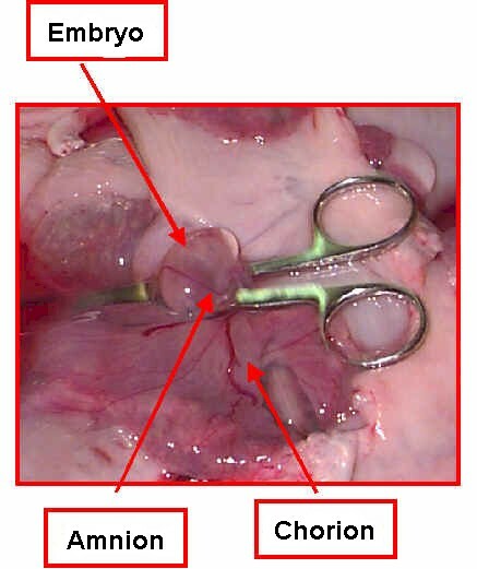
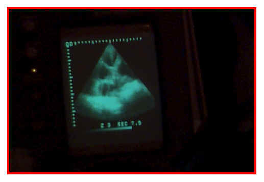
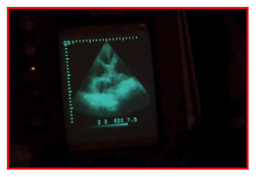
By days 25-35, the embryo is about 1 to 1 1/2 inches long, and major body systems and appendages are well formed. Each embryo is surrounded by a separate series of fluid-filled membranes, the amnion and chorion, which are comprise the placenta. These membranes help to protect and nourish the growing embryo. Nutrients, waste, gases and certain antibodies cross the membranes between the dam and embryos blood systems. It is the presence or absence of this fluid that is detected by the commonly used "ultrasound" pregnancy diagnosis equipment.
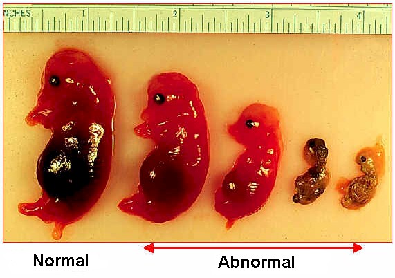
The fetal period begins at approximately day 36. Sexes may be easily determined by external examination and the main systems of the body are more well defined. The fetus can now be called a miniature adult. Fetal orientation is random; some are head to head, some are tail to tail and some are head to tail. At farrowing, about half are born tail first and half are born head first. Embryos which die before day 35-40 are usually reabsorbed by the dam. However, progressive calcification of the skeleton begins to occur from day 36 onward and deaths occurring after this point will result in mummification.

By day 109 the fetus weighs about 1½ to 2 pounds. Hair shafts begin to emerge but remain trapped under a skin layer until close to birth. Throughout gestation, the uterus gradually enlarges from about 2 to 3 pounds at mating to up to 60 pounds, including fetal contents, during the last week. Some females will lose up to 10 to 11% of their body weight at parturition or farrowing.
The average gestation length or time from conception until farrowing is 114 days. During gestation, the piglet grows from the union of a microscopic sperm and ovum into a fully formed individual weighing from 3 to 3½ pounds.
