AHVLA Pig Disease Surveillance Monthly Report: April 2013
Highlights from this report from the AHVLA include Porcine Respiratory and Reproductive Syndrome (PRRS) underlying post-weaning disease, Glässers disease prominent with concurrent salmonellosis and an unusual neonatal disease suspected to be due to Mycoplasma suis infection. 1 September 2013
 9 minute read
 By:
By: Alimentary Disease
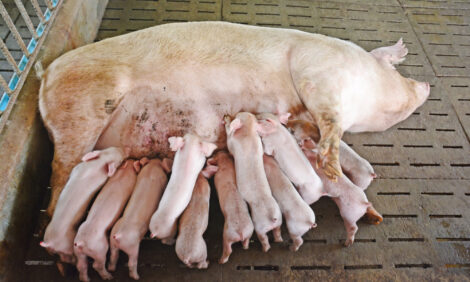
Multiple enteropathogens in pre-weaned pigs with ill-thrift
A small farrow-to-finish unit with 15 sows had a problem where two to three piglets in each litter started to fade at about four weeks of age prior to weaning at eight-weeks-old. Affected piglets became hairy and some had diarrhoea.
Three live eight-week-old piglets in poor body condition and with diarrhoea were submitted to Thirsk for investigation.
There were milk spots in the liver indicating migration of Ascaris suum. There was enterocolitis and histology and virology revealed evidence of rotavirus and coccidiosis. The advice was to instigate routine early treatment for coccidiosis and a worming programme.
Respiratory Disease
Mixed respiratory disease in growers found dead
Six eight-week-old pigs were found dead on one day from a group of 400 growing pigs and two were submitted to Penrith. The consistent finding in the animals was a fibrinous polyserositis and multifocal pneumonia.
Actinobacillus pleuropneumoniae and Bordetella bronchiseptica were isolated from the lung of one pig, while Streptococcus suis type 2 was isolated from the lung of another pig. Although coincidental that the pigs were found dead together, it appears that there were two distinct causes of death, although precipitating factors including ventilation or other environmental stresses may have been similar.
Complex respiratory disease in finishers
Two fresh plucks were submitted from an indoor finisher unit operated all-in all-out on a shed basis. Approximately 20 per cent of 600 18-week-old finishers were coughing, dyspnoeic and lethargic with unevenness; 20 had died, 10 in the week prior to submission. There was some response to penicillin by injection. Pigs were vaccinated for PCV2 and Mycoplasma hyopneumoniae.
In one pluck, there was cranioventral poorly demarcated consolidation from which no bacteria were isolated but Mycoplasma hyopneumoniae was detected and some histopathological lesions supported mycoplasmal involvement.
In the second pluck, there was clear cranioventral pneumonia affecting all lung lobes from which Pasteurella multocida was isolated, together with Streptococcus dysgalactiae subspecies equisimilis causing a vegetative valvular endocarditis. This organism is seen occasionally, causing septicaemia and/or suppurative conditions in pigs. Lung histopathology on this pluck supported the involvement of Pasteurella multocida in the pneumonia with little evidence of mycoplasmal involvement.
High morbidity respiratory disease likely due to Glässer’s disease
Two dead 10-week-old pigs in good body condition were submitted to investigate respiratory disease occurring three weeks post-weaning on a nursery unit in which 1,500 of 2,500 pigs were reported to be affected with 25 deaths.
On-farm post mortem examination was suggestive of Glässer’s disease and the fibrinopurulent pleurisy, pericarditis and peritonitis in the two pigs submitted was also consistent with Glässer’s disease.
No bacteria were isolated from multiple sites to confirm the diagnosis although the absence of any other bacterial growth supported the likely diagnosis of Glässer’s disease; no viral involvement was detected.
Occasionally, it is not possible to isolate Haemophilus parasuis from suspect cases, even if there has been no recent antimicrobial treatment, as the organism is fastidious. Sampling multiple sites from multiple recently dead acute untreated clinical cases increases the likelihood of isolating the organism.
PRRS underlying polyserositis and salmonellosis in weaners
Two live and two dead seven-week-old pigs were submitted to Thirsk for postmortem examination to investigate coughing and wasting affecting a group of 500 pigs on 900-sow indoor farrow-to-finish unit. The unit was PRRS- and EP-positive and had recently instituted a programme of swine influenza vaccination of the breeding herd to control influenza diagnosed previously in weaners.
Postmortem examination revealed widespread polyserositis with arthritis. In three of the four pigs there was also diphtheritic typhlocolitis with yellow flocculent diarrhoea. Salmonella 4,12:-: (monophasic Salmonella Typhimurium) was isolated these pigs’ faeces together with a Streptococcus suis type 1/2 from the lung of one pig.
Significantly, PRRS genotype 1 (European strain) was detected by PCR in pooled sera from the four pigs indicating an active PRRSv challenge. Segregation between batches is reportedly very good on this farm and subsequent batches were affected to a much lesser extent. As a result of the laboratory findings, the PRRS vaccination protocol in the sows and incoming gilts was amended in an attempt to stabilise herd immunity.
Systemic Disease
Management and hygiene issues underlying disease in preweaned pigs
Three live pigs were submitted to investigate a problem of lethargy, poor growth, sneezing and coughing in approximately 15 per cent of 800 pre-weaned pigs around 23-days-old with a minimal increase in mortality.
Only one of the pigs submitted had respiratory disease with cranio-ventral pulmonary consolidation while the other two had significant oral lesions affecting the gingivae around fractured clipped teeth and swollen joints due to polyarthritis. Two of the pigs also had small umbilical abscesses. Haemophilus parasuis was isolated from the joint of one pig and, although not detected in the lung, it is possible that H. parasuis was involved in the respiratory disease on the unit. No swine influenza or PRRS viruses were detected.
Management and hygiene issues raised by the findings in the submitted pigs were immediately addressed by the attending veterinary surgeon.
Glässer’s disease with salmonellosis on two rearing units
On the first of these units, disease was diagnosed together with PRRS. Four live pigs were submitted from an outdoor rearing unit to investigate diarrhoea and ill-thrift affecting approximately 250 of 380 eight-week-old pigs from which 20 had died since weaning, despite antimicrobial treatment. One of the submitted pigs had a generalised fibrinous polyserositis suggestive of Glässer’s disease which was confirmed by isolation of Haemophilus parasuis and, in this pig, PRRS virus was also detected by PCR on the spleen. In the other three pigs, there was diarrhoea with thickening but no, or minimal, necrosis of the large intestine, and salmonellosis was confirmed by isolation of Salmonella Typhimurium phage type U288. No PRRS virus was detected in these pigs. As one of the pigs had dysentery, Brachyspira testing was undertaken with none detected. PRRS virus was only detected in the pig with Glässer’s disease, and as the pigs were vaccinated at five-weeks-old and submitted at eight-weeks-old, this could have represented vaccinal virus.
To further investigate PRRSv involvement, immunohistochemistry was undertaken on the lung of this pig. This confirmed PRRS virus involvement in the pneumonia, indicating that the PRRSv infection was clinically significant and, thus, likely to represent field challenge. Analysis of the PRRS virus involved is in progress.
In the second unit, there was complex disease involving Glässer’s disease with salmonellosis and rotaviral enteritis in five-week-old pigs submitted to investigate weight loss, swollen joints, some scour, recumbency and some sudden deaths. Four hundred and eighty of 800 five-week-old pigs were described as affected with a total of 20 having died since entry from a single source. A poor response to colistin treatment was reported.
The submitted pigs were found dead and Haemophilus parasuis was isolated from at least one site in all three pigs, one of which had a fibrinous polyserositis typical of Glässer’s disease and the other two had H. parasuis septicaemias and polyarthritis. Two of the pigs also had diarrhoea, and salmonellosis was diagnosed: a monophasic Salmonella Typhimurium-like organism was isolated (phage type 193) along with concurrent rotaviral infection.
In view of the reported poor response to colistin treatment, colistin minimum inhibitory concentration (MIC) was performed and the salmonella isolated was sensitive. The poor response may have reflected insufficient intake of medicated water.
Unusual neonatal disease suspected to be due to Mycoplasma suis infection
Pigs suspected to have been stillborn were submitted to Bury St Edmunds from a large indoor breeder-finisher with an ongoing problem of increased stillbirths affecting around 20 per cent of litters. All parities were affected and sows remained healthy. Some pigs were seen to be lethargic and pale at birth, then improving.
Nine dead pigs were submitted from three litters and, of these, only two were true stillbirths; the others had breathed and walked, and two had ingested colostrum.
No infectious cause of the problem was diagnosed from this submission; however, due to its similarity with a previous case of in utero infection with Mycoplasma suis, EDTA bloods were submitted from several typically affected piglets, and M. suis was detected in anaemic pigs by DGGE/PCR. In utero transmission of M. suis has been associated with neonatal disease and also with dysgalactia in sows.
Further information is in the following link and publications:
- http://www.defra.gov.uk/ahvla-en/files/pub-survrep-p0112.pdf
- Strait and others (2012) Journal of the American Veterinary Medical Association. 241:1666-1667
- Henderson and others (1997) Veterinary Record. 140:144-146.
Strategic medication of sows with tetracycline and avoidance of any potential iatrogenic transmission, for example by avoiding reuse of needles, are interventions to consider.
Streptococcal disease causing deaths of well-grown pre-weaned pigs
Two dead three-week-old pigs from one litter were submitted to Bury St Edmunds in good body condition having been found dead on an outdoor breeding herd. Several other litters in the same farrowing batch had piglets showing malaise, lameness and deaths over two to three days with up to half of the litter affected.
Both pigs submitted had non-specific gross lesions including fibrin stranding in the peritoneal cavity, excess fibrinous pericardial and pleural fluid and petechiation on the epicardium. Streptococcus suis type 1 was isolated in pure growth from viscera and joints.
The clinical history and gross lesions did not distinguish this outbreak of pre-weaning streptococcal disease from the Klebsiella pneumoniae ssp pneumoniae septicaemias seen in the summer months of 2011 and 2012, and submission of similar cases is important to determine the aetiology.
More information is available at http://www.defra.gov.uk/ahvla-en/files/pub-vet-klebsiella.pdf.
Haemorrhagic disease in piglets due to isoimmune thrombocytopaenia
Thrombocytopaenia purpura was diagnosed at Bury St Edmunds as the cause of widespread haemorrhages and deaths in two-week-old pigs. The sows of affected litters were healthy and non-pyrexic and it was reported that one litter was primarily affected.
Generalised haemorrhages affecting a number of organs and systems including lymphoid tissue were found and prompted discussion with the submitting farmer and veterinarian to determine whether there were grounds to suspect swine fever. Based on further history received, swine fever was not suspected and further testing supported isoimmune thrombocytopaenia as the most likely cause of the multisystemic haemorrhages, anaemia and low platelet counts.
This can occur if the sow produces antibodies to foetal thrombocyte antigens, when the piglets absorb these antibodies from colostrum they damage platelets resulting in failure of blood clotting mechanisms.
Deaths and nervous signs due to Streptococcus suis 2 infection
Two dead pigs were submitted from a nursery-finisher with a history of mortality post-weaning, with Streptococcus suis and swine influenza diagnosed in a previous submission. The current batch had received antimicrobial medication in the water until two weeks prior to submission; 15 pigs out of 900 were either found dead or seen to have nervous signs since medication finished.
Non-specific gross lesions were found and Streptococcus suis type 2 was isolated from the meninges and lung, confirming meningitis and septicaemia due to streptococcal disease, on this occasion without swine influenza.
September 2013