Emerging Threats Quarterly Report – Pig Diseases - January-March 2013
Among the highlights of the latest quarterly surveillance report from AHVLA are testing of Strep suis for penicillin sensitivity, Border disease in a commercial herd and severe outbreaks of erysipelas. 31 July 2013
 13 minute read
 By:
By: Highlights
These reports aim to identify emerging animal disease related threats. Their production is underpinned by a large amount of surveillance data and information compiled as part of the Defra Food and Farming Group animal disease surveillance programme. Some of these data can be viewed on the AHVLA website.
- Penicillin sensitivity testing of Streptococcus suis
- Border disease virus infection diagnosed in a commercial pig herd
- Severe skin haemorrhages in weaners with erysipelas
- Serotyping of Erysipelothrix species isolates from diseased pigs
Ongoing Emergency Disease Investigations
Penicillin sensitivity testing in Streptococcus suis isolated from pigs in England
The detection of two 2009-10 clinical isolates of Streptococcus suis from pigs with raised penicillin Minimum Inhibitory Concentration (MIC) values above the human clinical breakpoint for central nervous system (CNS) infection, was described in the October to December 'Emerging Threats' report.
This has been investigated further by MIC testing of all clinical S. suis isolates from 2011 to 2013. None of these 215 isolates had MIC values above the human clinical breakpoint (>0.25mg/l) for central nervous system (CNS) infection. Most had penicillin MIC values between 0.006 and 0.064 mg/l while four isolates had slightly higher MIC values of 0.125 mg/l. These four were from 2011, 2012 and 2013 and three were serotype 2, which was the most common serotype tested. In March 2013, a clinical isolate of S. suis serotype 1 from the lung of a pig was found to be resistant to penicillin by routine disc diffusion antimicrobial sensitivity testing and this was confirmed by an MIC of 1.5 mg/l.
There is limited published data available on which to interpret these antimicrobial sensitivity findings. The current medical (BSAC) guidelines do not provide specific values for S. suis but give a clinical breakpoint in man of resistant >0.25mg/l for beta-haemolytic streptococci. The isolate is therefore resistant by those criteria and the MIC should be considered as significantly raised at least. It is possible that at this MIC, infection of viscera (for example, lung) with this organism may respond to penicillin, but, because of the blood brain barrier and pharmacokinetic and pharmacodynamic characteristics of the antimicrobial, that CNS infections may not.
This S. suis type 1 was considered likely to be playing a secondary role to Actinobacillus pleuropneumoniae in the clinical disease in the pig. The testing of recent AHVLA S. suis isolates has not provided evidence of continuing emergence of penicillin resistance but the recent detection of one resistant 2013 isolate highlights the need to maintain surveillance. MIC testing of AHVLA S. suis isolates will be undertaken to monitor the situation.
Border disease virus infection diagnosed in a commercial pig herd
The cause of haemorrhagic disease resembling swine fever in a commercial herd described in the July to September 2012 Emerging Threats report (page 4 http://www.defra.gov.uk/ahvla-en/files/pub-survrepp0312. pdf) was established as Border Disease.
The Border Disease virus (BDV) was initially detected by the virus microarray and confirmed by specific PCR testing and gene sequencing. Further cases of illthrift and anaemia due to BDV infection in postweaned pigs were confirmed on the farm in March 2013 indicating continuing BDV infection in the herd over a period of at least nine months.
BDV immunohistochemistry on their brains revealed patterns of labeling very similar to those observed in persistent pestiviral infections in ruminants that have specific immunotolerance to pestivirus as a consequence of early gestational pestivirus infection, confirming the suspicion that the pigs were likely to have been infected in utero.
A farm visit was made to the mixed livestock holding comprising an indoor breeder-finisher and a flock of breeding ewes. Sheep handling facilities were immediately adjacent to the dry sow yard with no separation of air space and shared staff. This will have provided the opportunity for transmission of BDV between sheep and pregnant sows resulting in birth of viraemic piglets which subsequently developed clinical disease in the post-weaning period.
Several management practices were identified which are likely to have favoured continued transmission and propagation of BDV in the pig herd without any further introduction of infection from sheep having been necessary. These practices included keeping in-pig gilts in the fattener shed with a shared scrape-through dung channel and using empty boar pens in the service house for growing pigs with illthrift. Advice was given to cease these practices and to ensure that the homebred replacement gilts and boars were exposed to likely persistently BDV-infected pigs well in advance of use for breeding.
It is worth noting that the clinical presentation of disease in this case is similar to that which might initially be seen if breeding pigs were infected with classical swine fever virus of low virulence. When the first case was submitted, disease had been ongoing for several weeks on farm, the possibility of swine fever was considered and ruled out on clinical grounds, in part due to the absence of disease in in-contact pigs. This case is not considered to represent a threat to other pig units and is not the first report of BDV infection in pigs (Paton and Done, 1994). The case illustrates the usefulness of new virus discovery methods in unusual cases and highlights a disease which might be reported as suspect swine fever. Publications will be progressed.
Klebsiella pneumoniae septicaemia in pre-weaned pigs
No cases of Klebsiella pneumoniae subspecies pneumoniae (Kpp) septicaemia have been diagnosed since August 2012, which is consistent with the seasonal pattern of this disease which has been restricted to the summer months.
Kpp detection from nasal cavities and tonsils of pigs submitted to AHVLA for diagnosis of disease unrelated to Kpp septicaemia continues and has been extended till September 2013 to give 12 months surveillance and to cover the summer months.
To date, 19 Kpp isolates from this extended surveillance have been tested and none is the outbreak-related strain, Kpp ST25, and none carries the small plasmid which is so far unique to the Kpp ST25 isolated from pigs. This plasmid has been sequenced and work continues to elucidate if any function can be ascribed to the plasmid genes. Genome sequencing of selected porcine Kpp strains is planned.
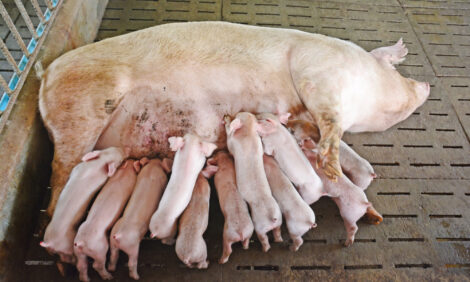
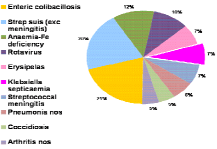
Interestingly, Kpp septicaemia was one of the 10 most frequent diagnoses in pre-weaned pigs in AHVLA submissions from 2011-12, representing seven per cent of diagnoses as illustrated in figure 1.
An oral presentation on Kpp septicaemia will be given to the joint Pig Veterinary Society-European Symposium of Porcine Health Management conference in May 2013. The re-emergence of Kpp septicaemia in 2012 was highlighted in quarterly and monthly reports and an information sheet is available: http://www.defra.gov.uk/ahvla-en/files/pub-vet-pfts.pdf
Unusual Diagnoses or Presentations
There were a number of unusual diagnoses this quarter; details of these have been included in monthly AHVLA reports and AHVLA highlights to BPEX, BPA and Pig Veterinary Society. These will be kept under review to assess whether they justify initiation of emerging disease investigations.
Mulberry heart disease diagnosed in pre-weaned piglets
Mulberry heart disease (MHD) was diagnosed by SAC in two-week-old piglets submitted from a farm reporting deaths mainly pre-weaning but also after weaning; some were sudden deaths while others died after being found recumbent with signs suspicious of meningitis but giving variable responses to antimicrobial treatment.
Post-mortem examination of two submitted piglets which were found dead revealed clear, serous effusions in the pleural and pericardial cavities and extensive haemorrhages throughout the myocardium. MHD was confirmed by histopathology.
A third piglet submitted had polyarthritis due to streptococcal infection. There was no evidence of meningitis in any of the piglets examined.
MHD is a very unusual diagnosis in suckling piglets. The farm was using home-mix rations for dry sows and lactating sows, with the required vitamin and mineral balancer added and it was recommended that the vitamin E and selenium content of sow diets be reviewed. The age incidence of MHD diagnoses at AHVLA and SAC will be kept under review to determine whether further cases occur in the pre-weaned age group.
Severe skin haemorrhages in weaners with erysipelas
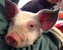
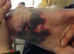
Unusually severe skin haemorrhages were reported and images of the lesions (figures 2 and 3) sent to AHVLA by a practitioner investigating disease in a group of 200 indoor weaners in which three pigs developed skin lesions four to seven days after weaning.
The affected pigs were bright and non-pyrexic; one died and two were euthanased and one was submitted to AHVLA where lesions were found consistent with a septicaemia but no significant organisms were isolated. However, Erysipelothrix species was isolated in pure and profuse growth from joint fluid of one of the other pigs with haemorrhagic skin lesions and kidney petechiation with no other remarkable lesions.
Within the same group, there were a few clinical cases of suspected acute erysipelas with pigs showing pyrexia, bright pink diamond skin lesions and the occasional enlarged hock joint and responding well to penicillin treatment. Other in-contact pigs and pigs sharing the same airspace were reported to be healthy and, across the farm, there was no evidence of other disease apart from minor coughing in the finishers and one case of porcine dermatitis and nephropathy syndrome in a group of finishers that had missed the usual PCV2 vaccination.
Although the skin lesions raised initial concern, the overall clinical picture indicated erysipelas as the likely cause and this was confirmed. The case was therefore not reported as suspect swine fever. The farm subsequently reported further cases of typical erysipelas in the same batch of weaners with no more haemorrhagic skin lesions. All affected pigs responded well to prompt penicillin treatment.
The inability to isolate Erysipelothrix species from the submitted case may be because the pig was euthanased and did not die of the disease and bacteraemia levels were too low for successful isolation.
This case highlights the importance of prompt veterinary attention and achieving a diagnosis in disease incidents, particularly where clinical signs resemble some of those seen in porcine notifiable disease.
Changes in Disease Patterns and Risk Factors
Serotyping of Erysipelothrix species isolates from outbreaks
Verbal reports from pig veterinarians of increased erysipelas outbreaks, some in replacement breeding pigs which should be fully vaccinated for erysipelas, were mentioned in the July to September 2012 'Emerging Threats' report (page 7 http://www.defra.gov.uk/ahvla-en/files/pub-survrep-p0312.pdf).
To assist in investigating whether this could relate to a change in serotype, AHVLA provided preparations of 99 historic and recent Erysipelothrix rhusiopathiae isolates from the AHVLA archive for serotyping which was kindly funded by MSD Animal Health UK. Forty-six have been successfully serotyped so far as indicated in Table 2 with more results to follow.
Interestingly, in the results received so far, serotype 1b has only been isolated since 2009 but serotyping results must be completed to allow full analysis and interpretation.
| Table 2. Serotypes of Erysipelothrix species isolates | ||||||
|---|---|---|---|---|---|---|
| Erysipelothrix Serotype | ||||||
| Year of isolation | Total | 11 | 2 | 1a | 1b | Untyped (to be repeated) |
| 1987 to 2013 | 99 | 4 | 27 | 12 | 3 | 53 |
| N.B. one serotype 11 isolate crossreacted with serotype 2 | ||||||
Table 3 shows the numbers of AHVLA and SAC erysipelas diagnoses in recent years. GB incidents of erysipelas were 3.3 per cent of diagnosable submissions so far for 2013 which is higher than in previous years although not significantly so, this will be kept under review.
| Table 3. Number of erysipelas diagnoses by year | ||||||
|---|---|---|---|---|---|---|
| Year | 2009 | 2010 | 2011 | 2012 | 2013* | |
| Number of diagnoses | 24 | 15 | 22 | 12 | 6* | |
| *period currently in progress | ||||||
Inclusion of details of outbreaks and this investigation in the quarterly and monthly reports has raised awareness and 2013 isolates will be included in the planned serotyping to investigate further.
Increased diagnoses of disease associated with Haemophilus parasuis
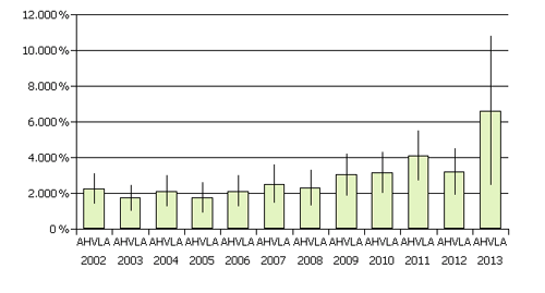
AHVLA diagnosed the highest rate of Haemophilus parasuis (Hps)-associated disease in the period January to March 2013 since 2002 as illustrated in Figure 4, in spite of the reduced carcass submission rate compared to the same period in 2012.
No cases were diagnosed by SAC. Outbreaks were diagnosed on eight units. Five of the diagnoses were of Glässer’s disease and three of Hps-associated pneumonia and six of the diagnoses were in the East Anglian region. All but one of the diagnoses were in postweaned pigs aged between five and 15 weeks old. Two diagnoses were of Glässer’s disease only, the other six were with a variety of systemic, enteric and respiratory diseases, in particular salmonellosis which was diagnosed on three occasions.
Details of these cases were included in AHVLA monthly reports for awareness and the trend will be assessed over the next quarter of 2013.
Swine influenza and PRRS disease incidents
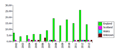
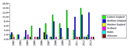
In the first quarter of 2012, increased GB diagnoses of swine influenza and PRRSv were reported compared to the same quarter in previous years. This increase was not been repeated in the first quarter of 2013 (Figure 5a), although there is a shift in regional distribution of diagnosed incidents with more being diagnosed in Northern England than in Eastern England, a reversal of the situation in the first quarter of 2012 (Figure 5b).
Horizon-scanning
Porcine circovirus-2 associated disease (PCVAD) in PCV-2 vaccinated pigs in North America
A recent publication detailed two outbreaks of clinical PCVAD on vaccinated pig farms in the United States in 2012 (Opriessnig and others, 2013). Both cases involved a novel PCV2b variant not previously detected in North America.
The two farms shared a common source of breeding gilts and disease occurred in 10– to 18-week-old pigs vaccinated against PCV2 . Both farms received replacement gilts from the same multiplier. The PCV2b strain identified was genetically very similar (99.9 per cent) to a recently described mutant PCV2 isolate reported in China and is considered to be more virulent than classical PCV2a or PCV2b strains.
The authors speculate that current PCV2a-based commercial vaccines may not be fully protective against this new strain. Recent AHVLA diagnoses of PCVAD have been in unvaccinated pigs. Future diagnoses of PCVAD will be reviewed regularly to determine whether they involve vaccinated pigs. There is very little information on the genetic types of PCV-2 in GB pigs and how much material is archived and available for analysis will be explored.
Mycoplasma bovis infection in Austrian pigs in contact with cattle
A publication highlighted the possibility of Mycoplasma bovis infection spreading from cattle to pigs where they are in direct contact (Spergser and others, 2013).
The paper describes an outbreak of disease due to M. bovis in cattle and pigs kept on the same mountain pasture in the Austrian Alps where the pigs were also fed with a diet containing whey from the affected dairy herd. The infected pigs developed severe conjunctivitis, nasal discharge and coughing.
This serves to raise awareness of another example of an pathogen of other farmed species which might cause disease in pigs on a mixed livestock enterprise.
References
Opriessnig T., Xiao C-T., Gerber P.F. and Halbur P.G. 2013. Emergence of a novel mutant PCV2b variant associated with clinical PCVAD in two vaccinated pig farms in the U.S. concurrently infected with PPV2. Veterinary Microbiology 163:177–183
Paton D.J. and Done S.H. 1994. Congenital infection of pigs with ruminant-type pestiviruses. Journal of Comparative Pathology 111:151-163
Spergser J, Macher K, Kargl M, Lysnyansky I, Rosengarten R. 2013. Emergence, re-emergence, spread and host species crossing of Mycoplasma bovis in the Austrian Alps caused by a single endemic strain. Vet. Microbiol. doi: Veterinary Microbiology 164:299-306. doi: 10.1016/j.vetmic.2013.02.007. Epub 2013 Feb 20
Further Reading
Find out more information on the diseases mentioned in this article by clicking here.
August 2013