Smaller Pig Producers Course 5: Greasy Pig Disease
For NADIS, Mark White, BVSc LLB DPM MRCVS, outlines the causes, prevention and treatment of this bacterial disease of the skin, typically in piglets and weaners. 20 June 2013
 7 minute read
 By:
By: Greasy Pig Disease is a bacterial infection of the skin of the pig, which is known by a variety of other names - Greasy Skin, Exudative Epidermitis or Marmite Disease. The primary cause of the disease is Staphylococcus hyicus, which is a common bacterium known to colonise the skin of many pigs without causing disease. As a group of diseases, it probably constitutes the most common skin ailment in pigs and is pathologically a dermatitis/epidermatitis, the most significant feature of which is that it is non-irritant, making the skin condition easy to differentiate clinically from the other common pig skin disease, sarcoptic mange, with which it can occur simultaneously.
There are a wide variety of presentations of the disease, ranging from localised lesions on the body, including the ear tips, to whole body effects. The disease can occur in all age groups from soon after birth up to adults and in the younger pig can be fatal.
Development of Disease
The causative organism lives on the skin surface of the pig, but will require some form of trigger mechanism to produce disease. This may be:-
- Damage to the skin, either from fighting, other disease (e.g. Sarcoptic Mange, Pig Pox, Pityriasis Rosea) or injuries from floors, feeders or pen divisions. Bird attacks can be a significant trigger in individual pigs.
- Scalding of the skin by chemicals or severe sunburn.
- A film of grease or faeces over the skin, under which the organism can multiply.
- High humidity levels, producing moisture on the skin, within which the organism can proliferate. These can exist outdoors as well as in indoor environments.
Staph hyicus can be found on the skin of most pig populations but there appears to be various strains of the bacteria and it is possible that new variants can be introduced (usually with incoming stock or pig contact) and cause outbreaks of disease.
Clinical Presentation
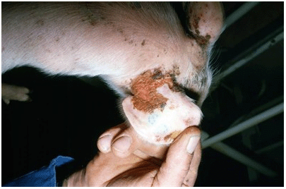
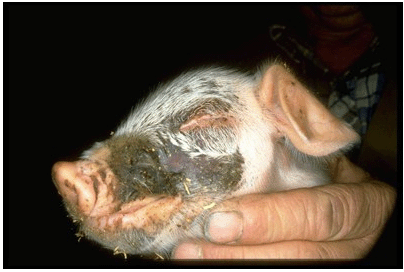
In the young pig, up to seven weeks of age, the most common presentation of Greasy Pig Disease is one of a brown to black development of scab appearance, starting usually around the shoulder and neck and spreading to part, or the whole, of the body. The younger it occurs, the more serious the consequences. It can occur in the sucking piglet as young as three to four days and, at this age, can be lethal due to the disturbance that occurs to fluid balance.
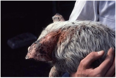
At the young end of the scale, an acute ulcerative dermatitis can occur, particularly on the soft skin of the abdomen and chest and can easily be mistaken for a contact ulceration, e.g. due to disinfectant or lime washing. This form of the disease can be seen as young as 24 hours old and is often fatal.
Occasionally, Staph hyicus can be involved in facial necrosis - the blackening of the skin of the face that results from teeth damage from litter mates where teeth clipping is not practised. (Fusobacterium necrophorum is the more common cause of this disease).
(although other bacteria are often involved)
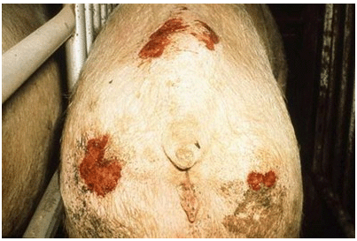
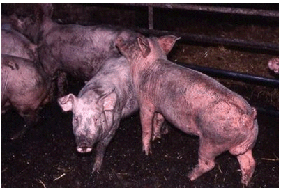
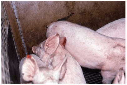
Moreover, discreet dermatitis will commonly occur around the head, neck and shoulders of weaned pigs as a result of colonisation of skin wounds - themselves the result of fighting - by staphylococci that are present on the skin. This form of the disease will often appear to spread throughout a group of weaners.
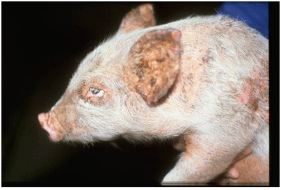
As older weaners and growers, the aftermath of earlier widespread greasy pig disease in individuals can be seen as part of the healing process. The skin will appear shiny, often hairless and have a distinct orange tinge to it. This healing process can be extremely slow, to the extent that it can still be unhealed by the time the pig reaches slaughter weight, resulting in condemnation of the skin.
likely to lead to skin condemnation and down-grading at slaughter
Staph hyicus is implicated in the development of dry gangrene of the extremities - affecting the tail of young piglets, often before tail docking has occurred and the ear tips of weaners of six to seven weeks upwards. The latter result is ear tip necrosis, an unsightly but apparently inherently harmless condition.
(ear tip necrosis)
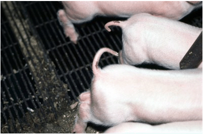
opens the door to Staph infection and tail necrosis
Localised greasy pig lesions can also occur on the legs of weaned pigs, starting at the foot and gradually creeping upwards. Here, primary damage occurs close to the coronary band of the hoof, allowing penetration of skin colonising bacteria.
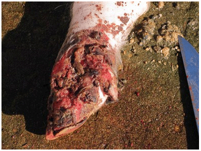
to damage to the coronary band
Finally, a rare and fatal form of Staph hyicus infection is reported in individual sows, in which the whole of the skin is affected, becomes progressively thickened and wrinkled (like rhinoceros skin) and progressive severe loss of condition occurs. These animals are usually euthanased.
Treatment
As a bacterial infection, Greasy Pig Disease responds well to antibiotic treatment, provided:
- the antibiotic used is effective against the strain involved
- sufficient dose is given for sufficient duration
- the antibiotic used penetrates into the skin in sufficient concentration, and
- treatment is given early.
Lincomycin, the penicillins and cephalosporins are most effective as injectable or orally applied treatments, although the latter should only be used as a last resort due to the implications of development of antibiotic resistance to these vitally important medicines in the human field.
Use of topical antibiotics direct onto the skin can be useful but there are no licensed products available in the UK for this purpose, although the advising veterinary surgeon may be able to prescribe "off licence" products under the cascade system.
For the young piglets, hydration is vital and any suckling pigs should be given electrolytes if Greasy Pig Disease occurs.
The use of soap (e.g. Savlon) can be a useful adjunct to treatment to remove the grease film over the skin and kill skin bacteria locally.
Any concomitant disease that is acting to trigger Greasy Pig Disease (e.g. sarcoptic mange) must also be treated.
Prevention
If any one feature can be highlighted that will precipitate development of lesions, it is skin damage, either as a result of fighting, abrasion by floors or injury on pen divisions etc, or scalding. Any control programmes must take this into account. Fighting can be reduced by minimising mixing and moving, maintaining stable groups and ensuring free access to feed, water and lying space. Reduced lighting can reduce aggression in the short term. Teeth clipping will reduce damage to skin from fighting (it should be done within the first three days of life subject to veterinary advice).
Fighting at weaning can be reduced by temporarily leaving pigs in the dark or spraying with a deodorant (e.g. Maskomal: Dupont), washing pigs in weak soap solution (e.g. Savlon) or disinfectant (e.g. Virkon S) prior to mixing. The latter techniques can reduce skin colonisation as well.
In addition, in the face of a clinical problem, many farms will wash pigs in Savlon or Virkon S (Dupont) to reduce skin contamination and thus reduce the risk of secondary infection with Staphylococcus hyicus into fight wounds. Care is needed not to chill pigs and to avoid excessive humidity whilst maintaining satisfactory airflow.
Where lesions start around the feet, attention to flooring is essential.
Whilst staphylococci are normal colonisers of the pigs' skin, the organism capable of producing Greasy Pig Disease can persist in the environment. Even in the smaller farms where there is a regular or consistent throughput in buildings or shelters, where disease is seen, a cleaning programme must be undertaken tailored to suit the structure. Wooden areas are difficult to clean and the use of creosote substitutes may be effective. Soaking in Virkon S at 1% dilution is also effective. High intensity ultraviolet light will be beneficial and as such, control may prove easier outdoors in summer.
In farms where long-standing intractable problems occur, use of strategic (in feed or in water) medication may be appropriate in anticipation of disease developing and, in some cases, autogenous or farm specific vaccines can be prepared to apply either to sows or to young pigs.
Conclusions
Staphylococcal dermatitis is common in pigs. It occurs at all ages in all different types of systems, producing a wide range of clinical presentations.
Early effective treatment is essential to relieve the suffering it causes and to avoid progression and spread.
Reducing trigger factors and attending to environmental hygiene are key to control.
June 2013