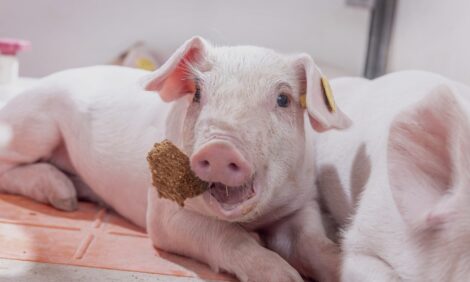
Gastric Ulcers: An Under-Recognized Cause of Mortality and Morbidity
By Robert M. Friendship; Department of Population Medicine, Ontario Veterinary College, University of Guelph - This paper, presented at the January 2003 Banff Pork Seminars, looks at the problem of gastric ulcers, a common cause of economic loss in pig production. 20 September 2003
 9 minute read

Introduction
Ulceration of the pars oesophagea is a common cause of death loss in the grower-finisher stage of pig production, and to a lesser extent, a cause of mortality in the sow herd. Stomach lesions can occur in glandular regions but these tend to be uncommon and generally associated with systemic diseases and will not be discussed in this paper.
The cause of pars oesophagea ulceration is not fully understood, but some of the important risk factors have been identified. Despite this relatively good level of understanding, the prevalence of gastric lesions at slaughter and the mortality rate due to gastric ulceration has increased over the past decade, and this problem has become one of the most important causes of death loss in the late finisher stage of production on many farms.
Prevalence and Severity of Ulcers
In slaughter house surveys, we typically find over 80% of market hogs with some type of lesion involving the pars oesophagea (Friendship, 1999). Instead of the smooth white glistening mucosal surface, the pars oesophagea more commonly has a roughened appearance (parakeratosis) and is often yellow in colour as a result of bile staining. Often, small erosions can be detected between the ridges when parakeratosis is present. The most common location for early erosions to occur is along the border between the pars oesophagea and the cardiac region of the stomach.
It is difficult to assess the severity of lesions on gross inspection alone. A small deep erosive lesion could result in severe blood loss, whereas an extensive lesion involving the entire pars oesophagea may be shallow and insignificant. Gastric ulcers are dynamic, occurring rapidly and possibly healing almost as fast. In addition to assessing the severity of an ulcer, it would be useful to be able to estimate the age of a lesion at slaughter, but this is very difficult.
Occasionally, the entire area of the pars oesophagea appears smaller than expected because of extensive scarring and sometimes it is absent, and the opening of the oesophagus into the stomach is restricted. In our studies, we commonly find pigs with oesophageal strictures at the opening of the stomach (Ayles et al,1996, Dionissopoulos et al, 2001). Sometimes the scarring is quite
severe making the opening very small and yet the pigs have grown relatively well with little clinical evidence of illness (possibly vomiting was observed).
Are Ulcers Related to Growth Rate?
Various researchers have examined the relationship between growth rate and stomach lesions at slaughter. Most studies have found that there is no relationship between gastric ulcer severity at slaughter and growth rate, unless lesions are severe and chronic (Friendship, 1999). Pigs with oesophageal strictures or anemia may be slower growing, but it is not uncommon to find severe gastric lesions in some of the fastest growing pigs. Unfortunately, an ulcer may have developed within the last day of the growing period, and other pigs with ulcers earlier in the growing stage may be healed at the time of slaughter. Studies have documented that when some pigs were slaughtered at arrival to the abattoir, they had less stomach ulcers than pigs from the same
barn that were held over night. We have tried to overcome this problem by monitoring pigs during the growing period using an endoscope, and noted an association between severity of stomach lesions and growth rate (Mackin et al., 1997).
I conclude from our studies that the major economic loss associated with gastric ulceration is mortality but I also believe that for every pig that dies, there are several other pigs in the same group that will have lesions that are severe enough to in some way interfere with growth performance. As a result of only considering mortality, we tend to underestimate the cost of gastric ulcers.
Feed Preparation and Ingredients
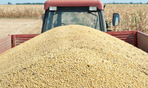
Feed has clearly an important role to play in the development of gastric ulcers. In North America, there has been a shift to feeding finely-ground, pelleted rations. If the particle size of the feed is small, the incidence of ulcer deaths increases, but the feed-to-gain efficiency improves. With our genotype of pigs and feed ingredients (corn and soybean meal), the point where ulcers begin to appear is when the mean particle size is less than about 700-750 microns.
Small feed particle size is associated with an increase in the fluidity of the stomach contents and a greater mixing. It is hypothesized that this situation allows pepsin and digestive acids to be in continual contact with the ulcer prone mucosa of the pars oesophagea. With a coarse diet fed ad-libitum, the pig maintains a pH gradient between the acidic pyloric area, where mixing occurs,
and the quiet, neutral pars oesophagea region. We have found that one of the most practical interventions is to put a group of pigs suffering ulcer losses onto a coarse ration for a one or two week period to allow healing (Ayles et al., 1996).
It would appear that an empty stomach is also an important risk factor for the development of gastric ulcers. Finely-ground feed increases gastric emptying and increases the likelihood of an empty stomach. We have experimented with the supplementation of melatonin, which appears to slow gastric emptying and possibly reduce the risk of ulcers (Ayles et al., 1999).
The most practical solution to preventing an empty stomach is to ensure there is no disruption in the feeding program. There are a number of reports that suggest stressful housing and shipping conditions are associated with ulcers, however, studies deliberately attempting to mimic stress such as injection of corticosteroids fail to produce ulcers in the pars oesophagea. It is possible that the main effect of so-called “stressful” conditions is the effect on feed intake.
Studies which increase stocking density but allow for access to feeders have not shown an increase in ulcers, but likely where feeder space is limited and there is a lot of mixing of pigs and high stocking density, there will be pigs with empty stomachs. In the southern United States, where summer temperatures are quite hot and feed consumption becomes a problem, there is a welldocumented rise in the incidence of ulcer deaths (Deen, 1993).
Concurrent Disease and Ulcers
There is also a relationship between concurrent disease and ulcers. It is frequently recognized that an outbreak of acute respiratory disease in a grower-finisher barn results in an increase in ulcers. We have performed an experiment in which one group of pigs was infected with PRRS and mycoplasma and another group was free of these diseases. The pigs were of similar genetics and raised in identical housing conditions. Pigs with respiratory disease had a higher prevalence and more severe gastric lesions than the disease-free pigs (Dionissopoulos et al, 2001). It may be that the increase in gastric ulcers, in this case, is caused by the pigs developing anorexia and an empty stomach, but it may also be associated with histamine release. Histamine is a powerful acid secretory stimulant. Injection of histamine to pigs results in ulceration of the pars oesophagea.
Acid Secretion and Ulcers
Reducing acid secretion to allow healing is generally considered an important part of therapy. We have examined some of the modern H+/K+-ATPase inhibitors such as lansoprazole and omeprazole (see Table 1). It would appear that omeprazole given at a dosage of 40 mg daily to grower pigs results in elevated gastric pH and reduces the occurrence of ulcers (Friendship et al., 2000, Melnichouk et al., 1999). This might be useful under certain circumstances such as a very valuable breeding animal during a period of high risk, however, these products are economically impractical for general use.
| Table 1: The effect of fasting and medication on stomach lesions and gastric pH of pigs |
|||
| Feeding | omeprazole | Gastric pH | Ulcers / total |
| Ad-libitum |
Control 20 mg |
3.3 3.9 |
1/5 0/5 |
| 24 h fast |
Control 20mg 40mg |
2.8 3.1 4.1 |
4/5 4/5 0/5 |
| 48h fast |
Control 40mg |
3.3 4.8 |
5/5 2/5 |
Alkaline salts (buffers) have been investigated as practical feed additives to help neutralize acidity in the stomach and improve morphology of the gastric mucosa. The use of 1% Na HCO3 and 1% KHCO3 have been shown to reduce stomach lesions (Wondra et al., 1995)
Genetics
There is a strong genetic component to gastric ulceration. The heritability of gastric ulcers has been estimated to be 0.52 (Berruecos and Robinson, 1972), which is similar to many carcass characteristics. Conceivably, we have tended to select animals that are more and more prone to gastric ulcers and we have moved to the use of feeds, which are ulcerogenic. In addition, it appears that if we accept the use of exogenous porcine somatotropin, the prevalence of ulcers will increase still further.
Are Ulcers Infectious?
There are researchers who believe that gastric ulcers in swine may have an infectious component similar to peptic ulcer disease of humans. Several types of spiral-shaped bacteria are found in pigs’ stomachs and the prevalence of these organisms is likely widespread in the general swine population.
Studies have reported from 10% to nearly 100% of pigs examined to be positive. Differences in prevalence may reflect the different sensitivity of the procedures used for detection as well as the true presence of the bacteria. These microorganisms are likely spread by a fecal-oral route and therefore housing/management conditions might influence the prevalence within a population.
The pathogenesis of an infectious cause of gastric ulceration has not been worked out for swine. Gnotobiotic pigs experimentally infected with specific swine Helicobacter organisms develop an inflammatory response similar to experimental infection with H. pylori, except the reaction is distributed mainly in the fundus compared to the cardia and antrum regions of H. pylori infected
pigs (Krakowka et al., 1995).
These researchers were unable to create ulcerative lesions in the pars oesophagea with experimental inoculation of Helicobacter organisms but were able to create lesions using fermentative organisms a such as Lactobacillus plus a high carbohydrate diet (Krakowka et
al., 1998). The glandular region tends to be where Helicobacter colonize so that if they play a role in ulceration, they must do this in an indirect way.
Evidence that these spiral organisms are associated with ulceration of the pars oesophagea is inconsistent. There are several studies that have found a higher incidence of ulcers in pigs with Helicobacter organisms compared to pigs without the organisms, but we have found no association (Melnichouk et al., 1999). If an association is established, there still remains the difficult job of demonstrating that the organism is a cause of disease and not merely an opportunist. In humans, therapy to eradicate the H. pylori and the subsequent success of preventing reoccurrence of ulcers helped to convince the medical community of the organisms relevance. We have undertaken small trials using antibiotics and acid secretion inhibitors but have not observed a difference in
ulcer prevalence. It is still too early to state for certain whether or not there is an infectious component to gastric ulceration in swine.
Conclusion
In conclusion, gastric ulceration will continue to be an important problem as the swine industry tries to balance the economic advantages of finely-ground pelleted feeds with the danger of creating ulcers. It is unlikely that the problem will be easily fixed by the addition of buffers or antibiotics to the feed. Careful attention to good feed manufacturing practices, prevention of respiratory disease, and assurance that pigs have continuous unrestricted access to feed and water will be important components of a gastric ulcer preventative program.
To read or print the PDF version of this paper (which includes the references) Click Here (6 pages, opens in new browser)
Source: Paper presented at the Banff Pork Seminar - January 2003, published September 2003