Study reveals big differences in ileitis vaccines
Results showed significantly fewer intestinal lesions were found in pigs vaccinated with Enterisol Ileitis. 28 November 2018
 2 minute read
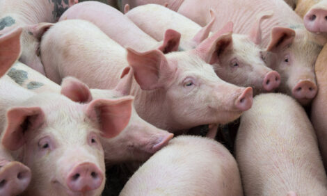
Most US hog farms are affected by ileitis, caused by the bacteria Lawsonia intracellularis. A recent study compared the efficacy of two ileitis vaccines, Enterisol® Ileitis and a competitor’s vaccine, by measuring and comparing intestinal lesions in weaned pigs challenged directly with Lawsonia after vaccination. [1] Previous research has established that increased prevalence and severity of ileocecal lesions correlates to decreased pig performance. [2]
Study results showed significantly fewer intestinal lesions were found in pigs vaccinated with Enterisol Ileitis than those vaccinated with the competitor’s vaccine or the control group.
Study method
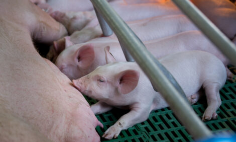
For the study, Boehringer Ingelheim (BI) technical veterinarians worked with researchers at Pipestone Applied Research and Pipestone Veterinary Services, Pipestone, Minnesota. Weaned pigs were placed in pens of 24, and pens were stratified by weight and gender, with an equal number placed in one of three treatment groups – one group vaccinated with Enterisol Ileitis orally at 8 weeks of age, another group vaccinated intramuscularly with a competitor’s vaccine at placement (3 weeks of age), and a third non-vaccinated control group. An equal number of non-vaccinated seeder pigs challenged with Lawsonia intracellularis were introduced to each group at 12 weeks of age. [1]
At four weeks post challenge, 30 pigs from each treatment group showing clinical signs of ileitis were randomly selected and necropsied, with tissues submitted to Iowa State University Veterinary Diagnostic Laboratory. Immunohistochemistry (IHC) and hematoxylin and eosin (H&E) staining of ileum and cecum sections were performed to detect the presence and severity of L. intracellularis and lesions resulting from bacterial colonisation and proliferation.
Study results
Microscopic lesions within the ileum sections were evident in all three treatment groups; however, the Enterisol Ileitis group had no lesions in the cecum section, and significantly fewer IHC positives (6.67 percent) in the ileum sections than the competitor vaccine’s group (43.33 percent) and the control group (63.33 percent). [1]
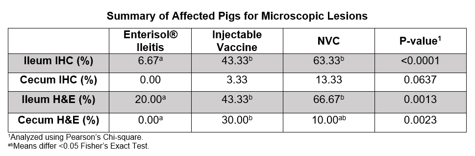
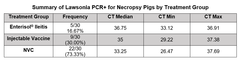
Both vaccinated groups had significantly smaller percentages of pigs affected in the ileum H&E sections than the non-vaccinated control group, but the Enterisol Ileitis group had fewer than half the pigs affected compared to the competitor vaccine’s group. The Enterisol Ileitis group also had lower numbers of Lawsonia PCR positives and lower levels from cecal content than the other two groups. Previous work has shown that more severe gut lesions and diarrhoea correlate to increased shedding of L. intracellularis. [3]
This study result showed that vaccinating with Enterisol Ileitis provides hogs with effective protection against ileitis, and it also minimises intestinal lesions, thereby helping to improve animal feed efficiency. However, if the time of post vaccination challenge is different, the results may not be the same.
References:
1 Dee S, Nerem J, Hanson D, et al. Comparison of intestinal lesions between Enterisol Ileitis and Porcilis Ileitis using a mucosal homogenate seeder pig challenge model, in Proceedings. AASV Annual Meeting; February 2017; 102–103.
2 Collins AM and Barchia IM. The critical threshold of Lawsonia intracellularis in pig feces that causes reduced average daily weight gains in experimentally challenged pigs. Vet Microbial 2014;168(2–4):455–458.
3 Burrough E, Rotolo M, Gauger PC, Schwartz KJ. Correlation of Lawsonia intracellularis semi-quantitative fecal polymerase chain reaction assay results with the presence of histologic lesions of proliferative enteropathy and positive immunohistochemical staining. J Swine Health Prod 2015;23(4):204–207.