

Comparing diagnostic sampling prospects from M. hyo-positive-source boar studs
Many sow farms have undergone Mycoplasma hyopneumoniae (M. hyo) elimination within their breeding herd, which makes it critical to understand the potential transmission risk from boar studs. While M. hyo is largely transmitted via nose-to-nose contact between pigs, systematic transmission has been speculated, and there has been limited documentation.Many sow farms have undergone Mycoplasma hyopneumoniae (M. hyo) elimination within their breeding herd, which makes it critical to understand the potential transmission risk from boar studs. While M. hyo is largely transmitted via nose-to-nose contact between pigs, systematic transmission has been speculated, and there has been limited documentation.
“There are only a few studies that have tested different diagnostic methods for M. hyo in boar studs or the transmission risk,” Zack Talbert, University of Illinois veterinary student, told Pig Health Today. Consequently, he wanted to dig deeper into the subject.
Talbert organized a study to evaluate four common diagnostic sampling techniques and assess the M. hyo transmission risk from a positive-source boar stud.1
The process
For the study, Talbert enrolled 90 boars from two studs known to previously receive M. hyo-positive replacement animals. These boars were sampled for M. hyo using four diagnostic sampling techniques:
- Tracheobronchial swabs (TBS) — This involved using the inner catheter of a post-cervical artificial insemination (PCAI) catheter, with an 80-mm FloqSwab attached at the end. After collection, the FloqSwab was removed and placed into a 3-mL tube containing 2 mL of phosphate buffered saline (PBS).
- Frothy oral fluids (FO) — These samples were collected using a 4 in. by 4 in. gauze square, secured onto the soft rubber end of a PCAI catheter. Before sampling, each modified PCAI catheter was dipped into a small Ziploc bag containing PBS. Froth was then swabbed from the boar’s mouth, after which the gauze square was placed into a small Ziploc bag and squeezed to release the fluid, which was transferred to a 3-mL tube.
- Semen (SEM) — 2 mL of fresh semen was placed in a 3-mL falcon tube prior to extending the semen.
- Serum (SM) — Lastly, 3 mL of blood was collected from the lateral saphenous vein.
All samples were sent to the Iowa State Veterinary Diagnostic Laboratory. Blood samples were tested by enzyme-linked immunosorbent assay (ELISA), and polymerase chain reaction (PCR) was conducted on the TBS, FO and SEM samples.
The results
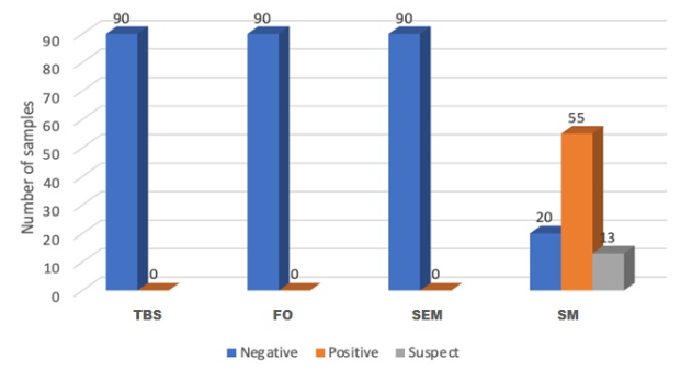
Both boar studs had seropositive animals identified by ELISA. Although positive ELISA tests were not surprising due to M. hyo vaccination protocols in isolation, several boars had an S:P ratio >1.0, indicating likely past exposure. All TBS, FO and SEM samples were PCR negative for M. hyo. (See accompanying chart.)

“Given the history of positive M. hyo-sourced boars into these studs, this study demonstrated that the transmission possibility may be minimal,” Talbert noted. More research is needed to validate the isolation of M. hyo SEM, he added.
Given the lack of research in this area, Talbert emphasized that those owning and managing boar studs should continue to be diligent in understanding lateral transmission prospects, elimination options and diagnostic testing methodology in order to assure classified health status.


