


4.4.8 PCV2
In the mid 1990’s in “association” with a new and distinct health problem of pigs called PMWS (post-weaning multi-systemic wasting syndrome) researchers at the University of Saskatchewan (Clark, Ellis, Harding, and West) identified a swine porcine circovirus (PCV2). Currently researchers from several different laboratories have demonstrated that PCV2 is a distinct virus (genetically and antigenically) in pigs causing PMWS. There is some evidence that PCV2 may be involved with several other swine health problems as well.
As of 2000, it has been reported across Canada, United States, and in many European (France, Spain, United Kingdom, Italy, Netherlands, Germany) and Asian countries. It is now seen as one of the most significant health problems in nursery and feeder pigs.
The strongest evidence for PCV2 involvement in swine disease is PMWS. This syndrome is seen primarily in pigs between 5 to 18 weeks of age.
Clinically it is characterized by “wasting” – pigs that fail to grow at the same rate as their pen-mates, then start to loose weight, eventually becoming extremely thin and emaciated. They may show signs of pneumonia and/or diarrhoea.
They are non-responsive to antibiotic therapy and, in spite of good treatment and extra care, continue to loose weight (see picture 4.4.8 a).
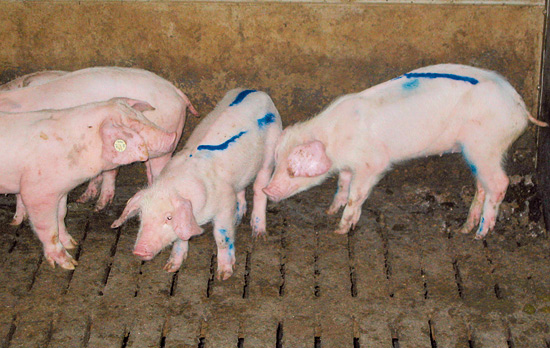
Picture 4.4.8 a (by R. Lising)
Group of pigs showing typical symptoms of PMWS.
The presence of PMWS varies tremendously from herd to herd; some herds appear to be disease free while in most herds that have had a positive case the number of pigs affected would be between 1 and 5%. In the few “outbreaks” that have been recorded, up to 50% of the pigs show symptoms and there is significantly elevated mortality.
Immunofluorescence tests and indirect or competitive ELISA’s are available to detect PCV2 specific antibodies. On post-mortem examination, these pigs are emaciated, many have stomach ulcers, and commonly there are signs of severe inflammation and/or degeneration in lung, kidney, liver and lymphoid tissue. Most striking are the enlarged inguinal and mesenteric lymphnodes (see picture 4.4.8 b).
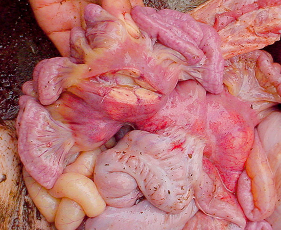
Picture 4.4.8 b (by R. Lising)
Enlarged mesenterial lymphnodes in a pig with PMWS.
PCV2-associated enteritis can be a distinct clinical manifestation of PCV2. Definitive diagnosis of porcine circovirus 2 (PCV2)-associated enteritis can be carried out by histopathology, virus isolation, and in situ hybridization. The most unique lesions are granulomatous inflammation affecting Peyer’s patches, characterized by infiltrates of epithelioid macrophages and giant multinucleated cells. Large, multiple, basophilic or amphophilic, grape-like intracytoplasmic inclusion bodies are often seen in the cytoplasm of histiocytic cells and giant multinucleated cells. The presence of diarrhoea and granulomatous enteritis, and abundant PCV2 DNA associated with the microscopic lesions is suggestive of PCV2-associated enteritis. PDNS (Porcine Dermatitis Nephritis Syndrome) (see picture 4.4.8 c) a complication which may occur in a PCV2 infection is of unknown aetiology.
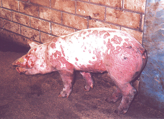
Picture 4.4.8 c (by H. Voets)
Fattening pig with typical signs of PDNS.
“Stress” is a significant factor in initiating the development of this disease in pigs. Concurrent diseases stress, nutritional stress, environmental stress, crowding, early weaning, poor sanitation, mixing pigs excessively, all can be factors that impact the development of this problem. The best plan to control and reduce PMWS – at this time is minimizing stress on the pigs where possible.
© Boehringer Ingelheim Animal Health GmbH, 2006
All rights reserved. No part of this Technical Manual 3.0 may be reproduced or transmitted in any form or by any means, electronic or photocopy, without permission in writing from Boehringer Ingelheim Animal Health GmbH.